2O4H

Human brain aspartoacylase complex with intermediate analog (N-phosphonomethyl-L-aspartate)


Experimental Data Snapshot

  • Method: X-RAY DIFFRACTION
  • Resolution: 2.70 Å
  • R-Value Free: 0.271 
  • R-Value Work: 0.202 
  • R-Value Observed: 0.205 

wwPDB Validation   3D Report Full Report


Ligand Structure Quality Assessment 


This is version 1.2 of the entry. See complete history


Literature

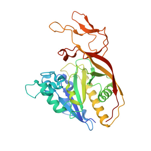
Examination of the Mechanism of Human Brain Aspartoacylase through the Binding of an Intermediate Analogue.

Le Coq, J.Pavlovsky, A.Malik, R.Sanishvili, R.Xu, C.Viola, R.E.

(2008) Biochemistry 47: 3484-3492

  • DOI: https://doi.org/10.1021/bi702400x
  • Primary Citation of Related Structures:  
    2O4H, 2O53

  • PubMed Abstract: 

    Canavan disease is a fatal neurological disorder caused by the malfunctioning of a single metabolic enzyme, aspartoacylase, that catalyzes the deacetylation of N-acetyl-L-aspartate to produce L-aspartate and acetate. The structure of human brain aspartoacylase has been determined in complex with a stable tetrahedral intermediate analogue, N-phosphonomethyl-L-aspartate. This potent inhibitor forms multiple interactions between each of its heteroatoms and the substrate binding groups arrayed within the active site. The binding of the catalytic intermediate analogue induces the conformational ordering of several substrate binding groups, thereby setting up the active site for catalysis. The highly ordered binding of this inhibitor has allowed assignments to be made for substrate binding groups and provides strong support for a carboxypeptidase-type mechanism for the hydrolysis of the amide bond of the substrate, N-acetyl- l-aspartate.


  • Organizational Affiliation

    Department of Chemistry, University of Toledo, Toledo, Ohio 43606, USA.


Macromolecules
Find similar proteins by:  (by identity cutoff)  |  3D Structure
Entity ID: 1
MoleculeChains Sequence LengthOrganismDetailsImage
Aspartoacylase
A, B
313Homo sapiensMutation(s): 0 
Gene Names: ASPAACY2ASP
EC: 3.5.1.15
UniProt & NIH Common Fund Data Resources
Find proteins for P45381 (Homo sapiens)
Explore P45381 
Go to UniProtKB:  P45381
PHAROS:  P45381
GTEx:  ENSG00000108381 
Entity Groups  
Sequence Clusters30% Identity50% Identity70% Identity90% Identity95% Identity100% Identity
UniProt GroupP45381
Sequence Annotations
Expand
  • Reference Sequence
Small Molecules
Experimental Data & Validation

Experimental Data

  • Method: X-RAY DIFFRACTION
  • Resolution: 2.70 Å
  • R-Value Free: 0.271 
  • R-Value Work: 0.202 
  • R-Value Observed: 0.205 
  • Space Group: P 42 21 2
Unit Cell:
Length ( Å )Angle ( ˚ )
a = 143.081α = 90
b = 143.081β = 90
c = 104.604γ = 90
Software Package:
Software NamePurpose
REFMACrefinement
MAR345dtbdata collection
HKL-2000data reduction
SCALEPACKdata scaling
REFMACphasing

Structure Validation

View Full Validation Report


Ligand Structure Quality Assessment 


Entry History 

Deposition Data

Revision History  (Full details and data files)

  • Version 1.0: 2007-11-06
    Type: Initial release
  • Version 1.1: 2011-07-13
    Changes: Advisory, Version format compliance
  • Version 1.2: 2023-08-30
    Changes: Data collection, Database references, Derived calculations, Refinement description