2O49

Crystal Structure of the N-terminal CUT domain of SATB1 Bound to Matrix Attachment Region DNA


Experimental Data Snapshot

  • Method: X-RAY DIFFRACTION
  • Resolution: 2.00 Å
  • R-Value Free: 0.255 
  • R-Value Work: 0.212 
  • R-Value Observed: 0.212 

wwPDB Validation   3D Report Full Report


This is version 1.2 of the entry. See complete history


Literature

Structural basis for recognition of the matrix attachment region of DNA by transcription factor SATB1.

Yamasaki, K.Akiba, T.Yamasaki, T.Harata, K.

(2007) Nucleic Acids Res 35: 5073-5084

  • DOI: https://doi.org/10.1093/nar/gkm504
  • Primary Citation of Related Structures:  
    2O49, 2O4A

  • PubMed Abstract: 

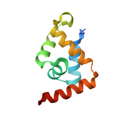
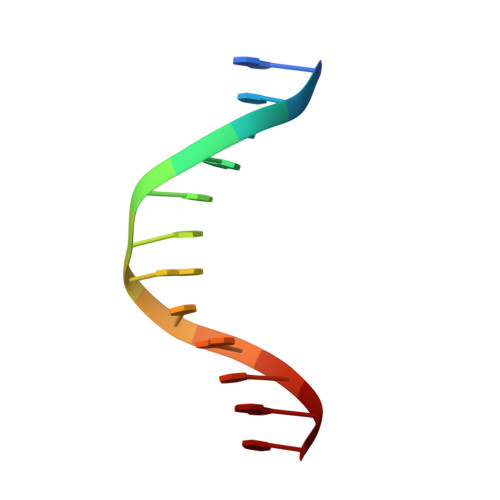
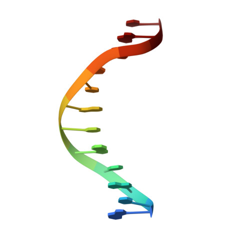
    Special AT-rich sequence binding protein 1 (SATB1) regulates gene expression essential in immune T-cell maturation and switching of fetal globin species, by binding to matrix attachment regions (MARs) of DNA and inducing a local chromatin remodeling. Previously we have revealed a five-helix structure of the N-terminal CUT domain, which is essentially the folded region in the MAR-binding domain, of human SATB1 by NMR. Here we determined crystal structure of the complex of the CUT domain and a MAR DNA, in which the third helix of the CUT domain deeply enters the major groove of DNA in the B-form. Bases of 5'-CTAATA-3' sequence are contacted by this helix, through direct and water-mediated hydrogen bonds and apolar and van der Waals contacts. Mutations at conserved base-contacting residues, Gln402 and Gly403, reduced the DNA-binding activity, which confirmed the importance of the observed interactions involving these residues. A significant number of equivalent contacts are observed also for typically four-helix POU-specific domains of POU-homologous proteins, indicating that these domains share a common framework of the DNA-binding mode, recognizing partially similar DNA sequences.


  • Organizational Affiliation

    Age Dimension Research Center, National Institute of Advanced Industrial Science and Technology (AIST), Tsukuba, Japan. yamasaki@aist.go.jp


Macromolecules

Find similar proteins by:  (by identity cutoff)  |  3D Structure
Entity ID: 3
MoleculeChains Sequence LengthOrganismDetailsImage
DNA-binding protein SATB1C [auth A]93Homo sapiensMutation(s): 0 
UniProt & NIH Common Fund Data Resources
Find proteins for Q01826 (Homo sapiens)
Explore Q01826 
Go to UniProtKB:  Q01826
PHAROS:  Q01826
GTEx:  ENSG00000182568 
Entity Groups  
Sequence Clusters30% Identity50% Identity70% Identity90% Identity95% Identity100% Identity
UniProt GroupQ01826
Sequence Annotations
Expand
  • Reference Sequence

Find similar nucleic acids by:  Sequence   |   3D Structure  

Entity ID: 1
MoleculeChains LengthOrganismImage
DNA (5'-D(*DGP*DCP*DTP*DAP*DAP*DTP*DAP*DTP*DAP*DTP*DGP*DC)-3')A [auth B]12N/A
Sequence Annotations
Expand
  • Reference Sequence

Find similar nucleic acids by:  Sequence   |   3D Structure  

Entity ID: 2
MoleculeChains LengthOrganismImage
DNA (5'-D(*DGP*DCP*DAP*DTP*DAP*DTP*DAP*DTP*DTP*DAP*DGP*DC)-3')B [auth C]12N/A
Sequence Annotations
Expand
  • Reference Sequence
Experimental Data & Validation

Experimental Data

  • Method: X-RAY DIFFRACTION
  • Resolution: 2.00 Å
  • R-Value Free: 0.255 
  • R-Value Work: 0.212 
  • R-Value Observed: 0.212 
  • Space Group: P 1
Unit Cell:
Length ( Å )Angle ( ˚ )
a = 29.659α = 71.6
b = 36.997β = 83.92
c = 41.248γ = 71.07
Software Package:
Software NamePurpose
SMARTdata collection
CNSrefinement
SAINTdata reduction
SCALAdata scaling
CNSphasing

Structure Validation

View Full Validation Report


Entry History 

Deposition Data

Revision History  (Full details and data files)

  • Version 1.0: 2007-08-14
    Type: Initial release
  • Version 1.1: 2011-07-13
    Changes: Version format compliance
  • Version 1.2: 2023-10-25
    Changes: Data collection, Database references, Refinement description