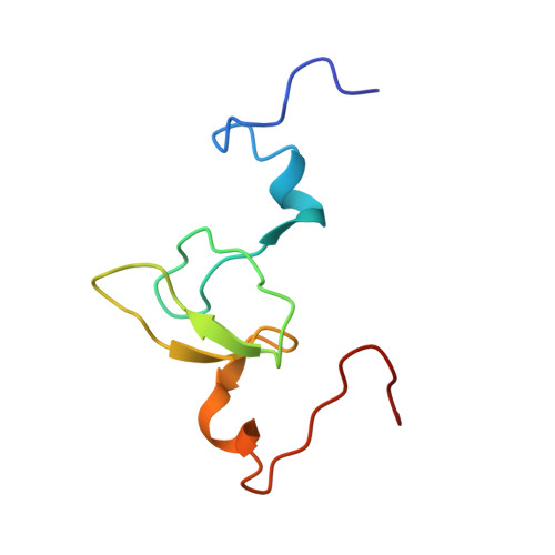
Structure of the HHARI catalytic domain shows glimpses of a HECT E3 ligase.
Spratt, D.E., Mercier, P., Shaw, G.S.(2013) PLoS One 8: e74047-e74047
- PubMed: 24058416
- DOI: https://doi.org/10.1371/journal.pone.0074047
- Primary Citation of Related Structures:
2M9Y - PubMed Abstract:
The ubiquitin-signaling pathway utilizes E1 activating, E2 conjugating, and E3 ligase enzymes to sequentially transfer the small modifier protein ubiquitin to a substrate protein. During the last step of this cascade different types of E3 ligases either act as scaffolds to recruit an E2 enzyme and substrate (RING), or form an ubiquitin-thioester intermediate prior to transferring ubiquitin to a substrate (HECT). The RING-inBetweenRING-RING (RBR) proteins constitute a unique group of E3 ubiquitin ligases that includes the Human Homologue of Drosophila Ariadne (HHARI). These E3 ligases are proposed to use a hybrid RING/HECT mechanism whereby the enzyme uses facets of both the RING and HECT enzymes to transfer ubiquitin to a substrate. We now present the solution structure of the HHARI RING2 domain, the key portion of this E3 ligase required for the RING/HECT hybrid mechanism. The structure shows the domain possesses two Zn²⁺-binding sites and a single exposed cysteine used for ubiquitin catalysis. A structural comparison of the RING2 domain with the HECT E3 ligase NEDD4 reveals a near mirror image of the cysteine and histidine residues in the catalytic site. Further, a tandem pair of aromatic residues exists near the C-terminus of the HHARI RING2 domain that is conserved in other RBR E3 ligases. One of these aromatic residues is remotely located from the catalytic site that is reminiscent of the location found in HECT E3 enzymes where it is used for ubiquitin catalysis. These observations provide an initial structural rationale for the RING/HECT hybrid mechanism for ubiquitination used by the RBR E3 ligases.
Organizational Affiliation:
Department of Biochemistry, Schulich School of Medicine and Dentistry, University of Western Ontario, London, Ontario, Canada.