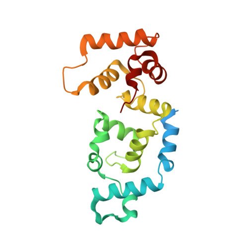
Structural basis for the activation of platelet integrin alphaIIb-beta3 by calcium- and integrin-binding protein 1.
Huang, H., Vogel, H.J.(2012) J Am Chem Soc 134: 3864-3872
- PubMed: 22283712
- DOI: https://doi.org/10.1021/ja2111306
- Primary Citation of Related Structures:
2LM5 - PubMed Abstract:
Calcium and integrin binding protein 1 (CIB1) is a specific binding partner for the cytoplasmic domain of the αIIb subunit of the highly abundant platelet integrin αIIbβ3. This protein has been suggested to be involved in the regulation of the activation of αIIbβ3, a process leading to platelet aggregation and blood coagulation. In this work, the solution structure of the deuterated Ca(2+)-CIB1 protein complexed with an αIIb peptide was first determined through modern RDC-based NMR methods. Next, we generated a complex structure for CIB1 and the αIIb domain (Ca(2+)-CIB1/αIIb) using the program Haddock, which is based on experimental restraints obtained for the protein-peptide interface from cross-saturation NMR experiments. In this data-driven complex structure, the N-terminal α-helix of the cytoplasmic domain of αIIb is buried in the hydrophobic pocket of the C-lobe of Ca(2+)-CIB1. The C-terminal acidic tail of αIIb remains unstructured and likely interacts with several positively charged residues in the N-lobe of Ca(2+)-CIB1. A potential molecular mechanism for the CIB1-mediated activation of the platelet integrin could be proposed on the basis of the model structure of this protein complex. Another feature of this work is that, in the NMR cross-saturation experiments, we applied the selective radio frequency irradiation to the smaller binding partner (the αIIb peptide), and successfully detected the binding interface on the larger binding partner Ca(2+)-CIB1 through its selectively protonated methyl groups. This 'reverse' methodology has a broad potential to be employed to many other complexes where synthetic peptides and a suitably isotope-labeled medium- to large-sized protein are used to study protein-protein interactions.
Organizational Affiliation:
Biochemistry Research Group, Department of Biological Sciences, University of Calgary, Calgary (AB), Canada, T2N 1N4.


