Structural basis of phosphoinositide binding to kindlin-2 protein pleckstrin homology domain in regulating integrin activation.
Liu, J., Fukuda, K., Xu, Z., Ma, Y.Q., Hirbawi, J., Mao, X., Wu, C., Plow, E.F., Qin, J.(2011) J Biol Chem 286: 43334-43342
- PubMed: 22030399
- DOI: https://doi.org/10.1074/jbc.M111.295352
- Primary Citation of Related Structures:
2LKO - PubMed Abstract:
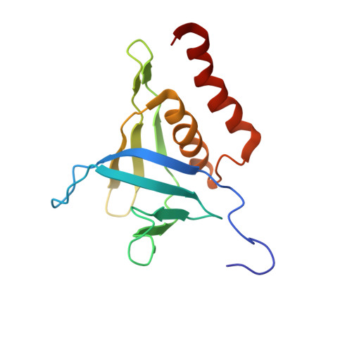
Kindlins are a subclass of FERM-containing proteins that have recently emerged as key regulators of integrin receptor activation and signaling. As compared with the conventional FERM domain, the kindlin FERM domain contains an inserted pleckstrin homology (PH) domain that recognizes membrane phosphoinositides, including phosphatidylinositol 4,5-bisphosphate (PIP2) and phosphatidylinositol 3,4,5-trisphosphate (PIP3). Using NMR spectroscopy, we show that PIP3 site-specifically binds to kindlin-2 PH with substantial chemical shift changes that are much larger than PIP2. This suggests an enhanced association of kindlin-2 with membrane as mediated by PIP3 upon its conversion from PIP2 by phosphoinositide-3 kinase, a known regulator of integrin activation. We determined the NMR structure of the kindlin-2 PH domain bound to the head group of PIP3, inositol 1,3,4,5-tetraphosphate (IP4). The structure reveals a canonical PH domain fold, yet with a distinct IP4 binding pocket that appears highly conserved for the kindlin family members. Functional experiments demonstrate that although wild type kindlin-2 is capable of cooperating with integrin activator talin to induce synergistic integrin α(IIb)β(3) activation, this ability is significantly impaired for a phosphoinositide binding-defective kindlin-2 mutant. These results define a specific PIP3 recognition mode for the kindlin PH domain. Moreover, they shed light upon a mechanism as to how the PH domain mediates membrane engagement of kindlin-2 to promote its binding to integrin and cooperation with talin for regulation of integrin activation.
Organizational Affiliation:
Department of Molecular Cardiology, Lerner Research Institute, Cleveland Clinic, Cleveland, Ohio 44195, USA.