Solution-state NMR structure and biophysical characterization of zinc-substituted rubredoxin B (Rv3250c) from Mycobacterium tuberculosis.
Buchko, G.W., Hewitt, S.N., Napuli, A.J., Van Voorhis, W.C., Myler, P.J.(2011) Acta Crystallogr Sect F Struct Biol Cryst Commun 67: 1148-1153
- PubMed: 21904065
- DOI: https://doi.org/10.1107/S1744309111008189
- Primary Citation of Related Structures:
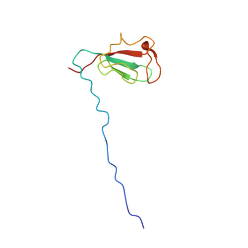
2KN9 - PubMed Abstract:
Owing to the evolution of multi-drug-resistant and extremely drug-resistant Mycobacterium tuberculosis strains, there is an urgent need to develop new antituberculosis strategies to prevent TB epidemics in the industrial world. Among the potential new drug targets are two small nonheme iron-binding proteins, rubredoxin A (Rv3251c) and rubredoxin B (Rv3250c), which are believed to play a role in electron-transfer processes. Here, the solution structure and biophysical properties of one of these two proteins, rubredoxin B (Mt-RubB), determined in the zinc-substituted form are reported. The zinc-substituted protein was prepared by expressing Mt-RubB in minimal medium containing excess zinc acetate. Size-exclusion chromatography and NMR spectroscopy indicated that Mt-RubB was a monomer in solution. The structure (PDB entry 2kn9) was generally similar to those of other rubredoxins, containing a three-stranded antiparallel β-sheet (β2-β1-β3) and a metal tetrahedrally coordinated to the S atoms of four cysteine residues (Cys9, Cys12, Cys42 and Cys45). The first pair of cysteine residues is at the C-terminal end of the first β-strand and the second pair of cysteine residues is towards the C-terminal end of the loop between β2 and β3. The structure shows the metal buried deeply within the protein, an observation that is supported by the inability to remove the metal with excess EDTA at room temperature. Circular dichroism spectroscopy shows that this stability extends to high temperature, with essentially no change being observed in the CD spectrum of Mt-RubB upon heating to 353 K.
Organizational Affiliation:
Seattle Structural Genomics Center for Infectious Disease, http://www.ssgcid.org, USA. garry.buchko@pnnl.gov