2JG9

Crystallographic structure of human C1q globular heads (P1)


Experimental Data Snapshot

  • Method: X-RAY DIFFRACTION
  • Resolution: 1.90 Å
  • R-Value Free: 0.260 
  • R-Value Work: 0.214 
  • R-Value Observed: 0.219 

wwPDB Validation   3D Report Full Report


This is version 1.4 of the entry. See complete history


Literature

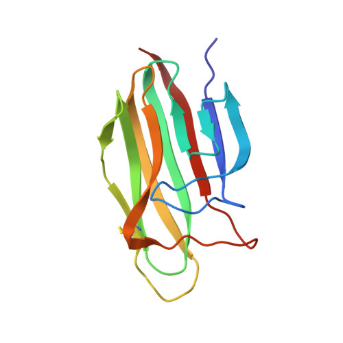
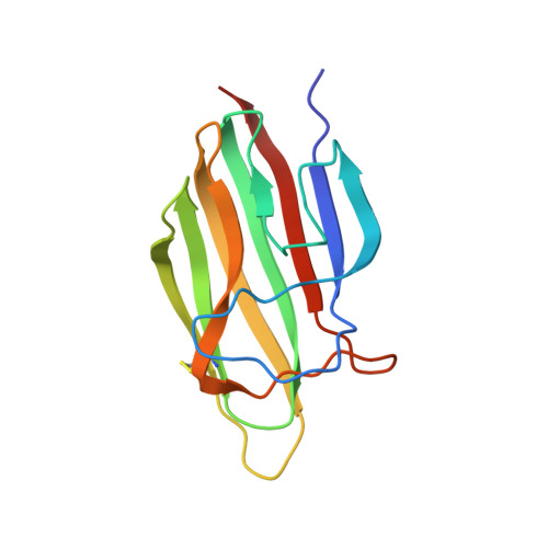
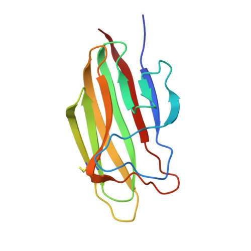
C1Q Binds Phosphatidylserine and Likely Acts as a Multiligand-Bridging Molecule in Apoptotic Cell Recognition.

Paidassi, H.Tacnet-Delorme, P.Garlatti, V.Darnault, C.Ghebrehiwet, B.Gaboriaud, C.Arlaud, G.J.Frachet, P.

(2008) J Immunol 180: 2329-2338

  • DOI: https://doi.org/10.4049/jimmunol.180.4.2329
  • Primary Citation of Related Structures:  
    2JG8, 2JG9

  • PubMed Abstract: 

    Efficient apoptotic cell clearance is critical for maintenance of tissue homeostasis, and to control the immune responses mediated by phagocytes. Little is known about the molecules that contribute "eat me" signals on the apoptotic cell surface. C1q, the recognition unit of the C1 complex of complement, also senses altered structures from self and is a major actor of immune tolerance. HeLa cells were rendered apoptotic by UV-B treatment and a variety of cellular and molecular approaches were used to investigate the nature of the target(s) recognized by C1q. Using surface plasmon resonance, C1q binding was shown to occur at early stages of apoptosis and to involve recognition of a cell membrane component. C1q binding and phosphatidylserine (PS) exposure, as measured by annexin V labeling, proceeded concomitantly, and annexin V inhibited C1q binding in a dose-dependent manner. As shown by cosedimentation, surface plasmon resonance, and x-ray crystallographic analyses, C1q recognized PS specifically and avidly (K(D) = 3.7-7 x 10(-8) M), through multiple interactions between its globular domain and the phosphoserine group of PS. Confocal microscopy revealed that the majority of the C1q molecules were distributed in membrane patches where they colocalized with PS. In summary, PS is one of the C1q ligands on apoptotic cells, and C1q-PS interaction takes place at early stages of apoptosis, in newly organized membrane patches. Given its versatile recognition properties, these data suggest that C1q has the unique ability to sense different markers which collectively would provide strong eat me signals, thereby allowing efficient apoptotic cell removal.


  • Organizational Affiliation

    Laboratoire d'Enzymologie Moléculaire, Institut de Biologie Structurale Jean-Pierre Ebel, 41 Rue Jules Horowitz, Grenoble Cedex 1, France.


Macromolecules
Find similar proteins by:  (by identity cutoff)  |  3D Structure
Entity ID: 1
MoleculeChains Sequence LengthOrganismDetailsImage
Complement C1q subcomponent subunit A
A, D
134Homo sapiensMutation(s): 0 
UniProt & NIH Common Fund Data Resources
Find proteins for P02745 (Homo sapiens)
Explore P02745 
Go to UniProtKB:  P02745
PHAROS:  P02745
GTEx:  ENSG00000173372 
Entity Groups  
Sequence Clusters30% Identity50% Identity70% Identity90% Identity95% Identity100% Identity
UniProt GroupP02745
Sequence Annotations
Expand
  • Reference Sequence
Find similar proteins by:  (by identity cutoff)  |  3D Structure
Entity ID: 2
MoleculeChains Sequence LengthOrganismDetailsImage
Complement C1q subcomponent subunit B
B, E
136Homo sapiensMutation(s): 0 
UniProt & NIH Common Fund Data Resources
Find proteins for P02746 (Homo sapiens)
Explore P02746 
Go to UniProtKB:  P02746
PHAROS:  P02746
Entity Groups  
Sequence Clusters30% Identity50% Identity70% Identity90% Identity95% Identity100% Identity
UniProt GroupP02746
Sequence Annotations
Expand
  • Reference Sequence
Find similar proteins by:  (by identity cutoff)  |  3D Structure
Entity ID: 3
MoleculeChains Sequence LengthOrganismDetailsImage
Complement C1q subcomponent subunit C
C, F
131Homo sapiensMutation(s): 0 
UniProt & NIH Common Fund Data Resources
Find proteins for P02747 (Homo sapiens)
Explore P02747 
Go to UniProtKB:  P02747
PHAROS:  P02747
GTEx:  ENSG00000159189 
Entity Groups  
Sequence Clusters30% Identity50% Identity70% Identity90% Identity95% Identity100% Identity
UniProt GroupP02747
Sequence Annotations
Expand
  • Reference Sequence
Experimental Data & Validation

Experimental Data

  • Method: X-RAY DIFFRACTION
  • Resolution: 1.90 Å
  • R-Value Free: 0.260 
  • R-Value Work: 0.214 
  • R-Value Observed: 0.219 
  • Space Group: P 1
Unit Cell:
Length ( Å )Angle ( ˚ )
a = 48.15α = 92.45
b = 48.24β = 92.65
c = 87.79γ = 113.53
Software Package:
Software NamePurpose
REFMACrefinement
XDSdata reduction
XSCALEdata scaling

Structure Validation

View Full Validation Report


Entry History 

Deposition Data

Revision History  (Full details and data files)

  • Version 1.0: 2008-02-19
    Type: Initial release
  • Version 1.1: 2011-05-08
    Changes: Version format compliance
  • Version 1.2: 2011-07-13
    Changes: Version format compliance
  • Version 1.3: 2018-12-05
    Changes: Data collection, Database references, Source and taxonomy, Structure summary
  • Version 1.4: 2023-12-13
    Changes: Data collection, Database references, Derived calculations, Other, Refinement description