2HNX

Crystal Structure of aP2


Experimental Data Snapshot

  • Method: X-RAY DIFFRACTION
  • Resolution: 1.50 Å
  • R-Value Free: 0.225 
  • R-Value Work: 0.174 
  • R-Value Observed: 0.177 

wwPDB Validation   3D Report Full Report


Ligand Structure Quality Assessment 


This is version 1.3 of the entry. See complete history


Literature

Expression, purification, crystallization and structure of human adipocyte lipid-binding protein (aP2).

Marr, E.Tardie, M.Carty, M.Brown Phillips, T.Wang, I.K.Soeller, W.Qiu, X.Karam, G.

(2006) Acta Crystallogr Sect F Struct Biol Cryst Commun 62: 1058-1060

  • DOI: https://doi.org/10.1107/S1744309106038656
  • Primary Citation of Related Structures:  
    2HNX

  • PubMed Abstract: 

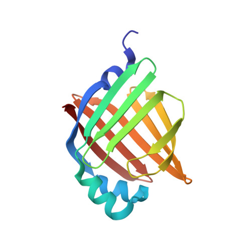
    Human adipocyte lipid-binding protein (aP2) belongs to a family of intracellular lipid-binding proteins involved in the transport and storage of lipids. Here, the crystal structure of human aP2 with a bound palmitate is described at 1.5 A resolution. Unlike the known crystal structure of murine aP2 in complex with palmitate, this structure shows that the fatty acid is in a folded conformation and that the loop containing Phe57 acts as a lid to regulate ligand binding by excluding solvent exposure to the central binding cavity.


  • Organizational Affiliation

    Exploratory Medicinal Sciences, Pfizer Global Research and Development Groton Laboratories, Eastern Point Road, Groton, CT 06340, USA.


Macromolecules
Find similar proteins by:  (by identity cutoff)  |  3D Structure
Entity ID: 1
MoleculeChains Sequence LengthOrganismDetailsImage
Fatty acid-binding protein, adipocyte136Homo sapiensMutation(s): 0 
Gene Names: FABP4
UniProt & NIH Common Fund Data Resources
Find proteins for P15090 (Homo sapiens)
Explore P15090 
Go to UniProtKB:  P15090
PHAROS:  P15090
GTEx:  ENSG00000170323 
Entity Groups  
Sequence Clusters30% Identity50% Identity70% Identity90% Identity95% Identity100% Identity
UniProt GroupP15090
Sequence Annotations
Expand
  • Reference Sequence
Experimental Data & Validation

Experimental Data

  • Method: X-RAY DIFFRACTION
  • Resolution: 1.50 Å
  • R-Value Free: 0.225 
  • R-Value Work: 0.174 
  • R-Value Observed: 0.177 
  • Space Group: P 21 21 21
Unit Cell:
Length ( Å )Angle ( ˚ )
a = 32.47α = 90
b = 53.94β = 90
c = 75γ = 90
Software Package:
Software NamePurpose
REFMACrefinement
HKL-2000data reduction
HKL-2000data scaling
AMoREphasing

Structure Validation

View Full Validation Report


Ligand Structure Quality Assessment 


Entry History 

Deposition Data

Revision History  (Full details and data files)

  • Version 1.0: 2006-11-28
    Type: Initial release
  • Version 1.1: 2008-05-01
    Changes: Version format compliance
  • Version 1.2: 2011-07-13
    Changes: Version format compliance
  • Version 1.3: 2024-02-14
    Changes: Data collection, Database references, Derived calculations