2HAV

Apo-Human Serum Transferrin (Glycosylated)


Experimental Data Snapshot

  • Method: X-RAY DIFFRACTION
  • Resolution: 2.70 Å
  • R-Value Free: 0.293 
  • R-Value Work: 0.232 
  • R-Value Observed: 0.232 

wwPDB Validation   3D Report Full Report


This is version 1.3 of the entry. See complete history


Literature

The Crystal Structure of Iron-free Human Serum Transferrin Provides Insight into Inter-lobe Communication and Receptor Binding.

Wally, J.Halbrooks, P.J.Vonrhein, C.Rould, M.A.Everse, S.J.Mason, A.B.Buchanan, S.K.

(2006) J Biol Chem 281: 24934-24944

  • DOI: https://doi.org/10.1074/jbc.M604592200
  • Primary Citation of Related Structures:  
    2HAU, 2HAV

  • PubMed Abstract: 

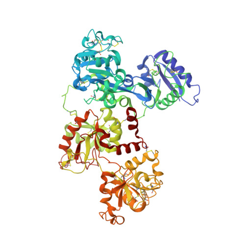
    Serum transferrin reversibly binds iron in each of two lobes and delivers it to cells by a receptor-mediated, pH-dependent process. The binding and release of iron result in a large conformational change in which two subdomains in each lobe close or open with a rigid twisting motion around a hinge. We report the structure of human serum transferrin (hTF) lacking iron (apo-hTF), which was independently determined by two methods: 1) the crystal structure of recombinant non-glycosylated apo-hTF was solved at 2.7-A resolution using a multiple wavelength anomalous dispersion phasing strategy, by substituting the nine methionines in hTF with selenomethionine and 2) the structure of glycosylated apo-hTF (isolated from serum) was determined to a resolution of 2.7A by molecular replacement using the human apo-N-lobe and the rabbit holo-C1-subdomain as search models. These two crystal structures are essentially identical. They represent the first published model for full-length human transferrin and reveal that, in contrast to family members (human lactoferrin and hen ovotransferrin), both lobes are almost equally open: 59.4 degrees and 49.5 degrees rotations are required to open the N- and C-lobes, respectively (compared with closed pig TF). Availability of this structure is critical to a complete understanding of the metal binding properties of each lobe of hTF; the apo-hTF structure suggests that differences in the hinge regions of the N- and C-lobes may influence the rates of iron binding and release. In addition, we evaluate potential interactions between apo-hTF and the human transferrin receptor.


  • Organizational Affiliation

    NIDDK, National Institutes of Health, Bethesda, Maryland 20892, USA.


Macromolecules
Find similar proteins by:  (by identity cutoff)  |  3D Structure
Entity ID: 1
MoleculeChains Sequence LengthOrganismDetailsImage
Serotransferrin
A, B
676Homo sapiensMutation(s): 0 
UniProt & NIH Common Fund Data Resources
Find proteins for P02787 (Homo sapiens)
Explore P02787 
Go to UniProtKB:  P02787
PHAROS:  P02787
GTEx:  ENSG00000091513 
Entity Groups  
Sequence Clusters30% Identity50% Identity70% Identity90% Identity95% Identity100% Identity
UniProt GroupP02787
Sequence Annotations
Expand
  • Reference Sequence
Small Molecules
Ligands 2 Unique
IDChains Name / Formula / InChI Key2D Diagram3D Interactions
CIT
Query on CIT

Download Ideal Coordinates CCD File 
C [auth A]
D [auth A]
E [auth A]
I [auth B]
J [auth B]
C [auth A],
D [auth A],
E [auth A],
I [auth B],
J [auth B],
K [auth B],
L [auth B]
CITRIC ACID
C6 H8 O7
KRKNYBCHXYNGOX-UHFFFAOYSA-N
GOL
Query on GOL

Download Ideal Coordinates CCD File 
F [auth A],
G [auth A],
H [auth A],
M [auth B]
GLYCEROL
C3 H8 O3
PEDCQBHIVMGVHV-UHFFFAOYSA-N
Experimental Data & Validation

Experimental Data

  • Method: X-RAY DIFFRACTION
  • Resolution: 2.70 Å
  • R-Value Free: 0.293 
  • R-Value Work: 0.232 
  • R-Value Observed: 0.232 
  • Space Group: P 21 21 21
Unit Cell:
Length ( Å )Angle ( ˚ )
a = 88.324α = 90
b = 103.258β = 90
c = 200.362γ = 90
Software Package:
Software NamePurpose
HKL-2000data reduction
PHASERphasing
REFMACrefinement
HKL-2000data scaling

Structure Validation

View Full Validation Report


Entry History 

Deposition Data

Revision History  (Full details and data files)

  • Version 1.0: 2006-06-27
    Type: Initial release
  • Version 1.1: 2008-05-01
    Changes: Version format compliance
  • Version 1.2: 2011-07-13
    Changes: Non-polymer description, Version format compliance
  • Version 1.3: 2023-08-30
    Changes: Data collection, Database references, Derived calculations, Refinement description