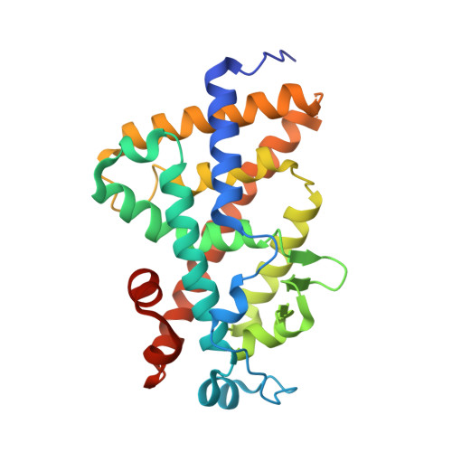
2HAS

Crystal structure of VDR LBD in complex with 2alpha-(1-propoxy) calcitriol


Experimental Data Snapshot

  • Method: X-RAY DIFFRACTION
  • Resolution: 1.96 Å
  • R-Value Free: 0.228 
  • R-Value Work: 0.186 
  • R-Value Observed: 0.188 

wwPDB Validation   3D Report Full Report


This is version 1.4 of the entry. See complete history


Literature

Probing a Water Channel near the A-Ring of Receptor-Bound 1alpha,25-Dihydroxyvitamin D3 with Selected 2alpha-Substituted Analogues

Hourai, S.Fujishima, T.Kittaka, A.Suhara, Y.Takayama, H.Rochel, N.Moras, D.

(2006) J Med Chem 49: 5199-5205

  • DOI: https://doi.org/10.1021/jm0604070
  • Primary Citation of Related Structures:  
    2HAM, 2HAR, 2HAS, 2HB7, 2HB8

  • PubMed Abstract: 

    The crystal structure of the vitamin D receptor (VDR) in complex with 1 alpha,25(OH)2D3 revealed the presence of several water molecules near the A-ring linking the ligand C-2 position to the protein surface. Here, we report the crystal structures of the human VDR ligand binding domain bound to selected C-2 alpha substituted analogues, namely, methyl, propyl, propoxy, hydroxypropyl, and hydroxypropoxy. These specific replacements do not modify the structure of the protein or the ligand, but with the exception of the methyl substituent, all analogues affect the presence and/or the location of the above water molecules. The integrity of the channel interactions and specific C-2 alpha analogue directed additional interactions correlate with the binding affinity of the ligands. In contrast, the resulting loss or gain of H-bonds does not reflect the magnitude of HL60 cell differentiation. Our overall findings highlight a rational approach to the design of more potent ligands by building in features revealed in the crystal structures.


  • Organizational Affiliation

    Laboratoire de Biologie et Génomique Structurales, UMR 7104, Institut de Génétique et de Biologie Moléculaire et Cellulaire, CNRS/INSERM/ULP, 1, Rue Laurent Fries, BP 10142, 67404 Illkirch Cedex, France.


Macromolecules
Find similar proteins by:  (by identity cutoff)  |  3D Structure
Entity ID: 1
MoleculeChains Sequence LengthOrganismDetailsImage
Vitamin D3 receptor263Homo sapiensMutation(s): 0 
UniProt & NIH Common Fund Data Resources
Find proteins for P11473 (Homo sapiens)
Explore P11473 
Go to UniProtKB:  P11473
PHAROS:  P11473
GTEx:  ENSG00000111424 
Entity Groups  
Sequence Clusters30% Identity50% Identity70% Identity90% Identity95% Identity100% Identity
UniProt GroupP11473
Sequence Annotations
Expand
  • Reference Sequence
Small Molecules
Ligands 1 Unique
IDChains Name / Formula / InChI Key2D Diagram3D Interactions
C3O
Query on C3O

Download Ideal Coordinates CCD File 
B [auth A]2ALPHA-PROPOXY-1ALPHA,25-DIHYDROXYVITAMIN D3
C30 H50 O4
DSWDAXGEZVIPHP-BRDKTQIRSA-N
Experimental Data & Validation

Experimental Data

  • Method: X-RAY DIFFRACTION
  • Resolution: 1.96 Å
  • R-Value Free: 0.228 
  • R-Value Work: 0.186 
  • R-Value Observed: 0.188 
  • Space Group: P 21 21 21
Unit Cell:
Length ( Å )Angle ( ˚ )
a = 45.1α = 90
b = 51.669β = 90
c = 131.715γ = 90
Software Package:
Software NamePurpose
ADSCdata collection
HKL-2000data reduction
CNSrefinement
HKL-2000data scaling
CNSphasing

Structure Validation

View Full Validation Report


Entry History 

Deposition Data

Revision History  (Full details and data files)

  • Version 1.0: 2006-08-29
    Type: Initial release
  • Version 1.1: 2008-05-01
    Changes: Version format compliance
  • Version 1.2: 2011-07-13
    Changes: Version format compliance
  • Version 1.3: 2017-08-16
    Changes: Refinement description, Source and taxonomy
  • Version 1.4: 2023-10-25
    Changes: Data collection, Database references, Derived calculations, Refinement description