2H9R

Docking and dimerization domain (D/D) of the regulatory subunit of the Type II-alpha cAMP-dependent protein kinase A associated with a Peptide derived from an A-kinase anchoring protein (AKAP)


Experimental Data Snapshot

  • Method: SOLUTION NMR
  • Conformers Calculated: 50 
  • Conformers Submitted: 10 
  • Selection Criteria: structures with the lowest energy 

wwPDB Validation   3D Report Full Report


This is version 1.3 of the entry. See complete history


Literature

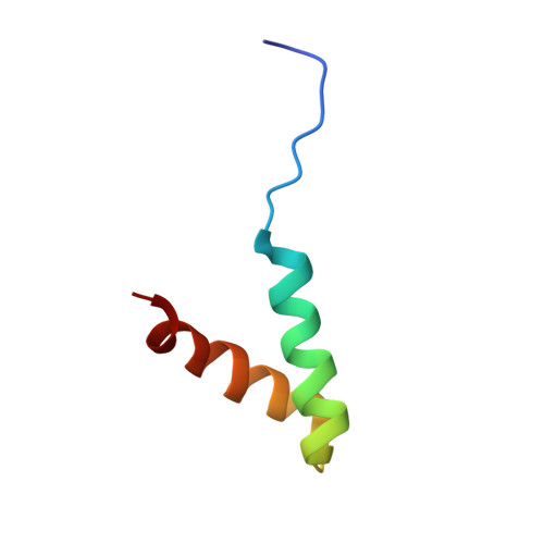
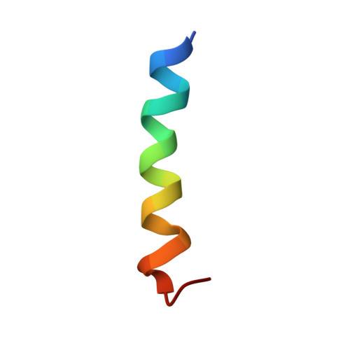
A novel mechanism of PKA anchoring revealed by solution structures of anchoring complexes.

Newlon, M.G.Roy, M.Morikis, D.Carr, D.W.Westphal, R.Scott, J.D.Jennings, P.A.

(2001) EMBO J 20: 1651-1662

  • DOI: https://doi.org/10.1093/emboj/20.7.1651
  • Primary Citation of Related Structures:  
    2DRN, 2H9R

  • PubMed Abstract: 

    The specificity of intracellular signaling events is controlled, in part, by compartmentalization of protein kinases and phosphatases. The subcellular localization of these enzymes is often maintained by protein- protein interactions. A prototypic example is the compartmentalization of the cAMP-dependent protein kinase (PKA) through its association with A-kinase anchoring proteins (AKAPs). A docking and dimerization domain (D/D) located within the first 45 residues of each regulatory (R) subunit protomer forms a high affinity binding site for its anchoring partner. We now report the structures of two D/D-AKAP peptide complexes obtained by solution NMR methods, one with Ht31(493-515) and the other with AKAP79(392-413). We present the first direct structural data demonstrating the helical nature of the peptides. The structures reveal conserved hydrophobic interaction surfaces on the helical AKAP peptides and the PKA R subunit, which are responsible for mediating the high affinity association in the complexes. In a departure from the dimer-dimer interactions seen in other X-type four-helix bundle dimeric proteins, our structures reveal a novel hydrophobic groove that accommodates one AKAP per RIIalpha D/D.


  • Organizational Affiliation

    The Department of Chemistry and Biochemistry, University of California, San Diego, La Jolla, CA 92093-0359, USA.


Macromolecules
Find similar proteins by:  (by identity cutoff)  |  3D Structure
Entity ID: 1
MoleculeChains Sequence LengthOrganismDetailsImage
cAMP-dependent protein kinase type II-alpha regulatory subunit
A, B
46Rattus norvegicusMutation(s): 0 
Gene Names: RIIA(1-44)
EC: 2.7.1.37
UniProt
Find proteins for P12368 (Rattus norvegicus)
Explore P12368 
Go to UniProtKB:  P12368
Entity Groups  
Sequence Clusters30% Identity50% Identity70% Identity90% Identity95% Identity100% Identity
UniProt GroupP12368
Sequence Annotations
Expand
  • Reference Sequence

Find similar proteins by:  Sequence   |   3D Structure  

Entity ID: 2
MoleculeChains Sequence LengthOrganismDetailsImage
22-mer from A-kinase anchor protein 522N/AMutation(s): 0 
UniProt & NIH Common Fund Data Resources
Find proteins for P24588 (Homo sapiens)
Explore P24588 
Go to UniProtKB:  P24588
PHAROS:  P24588
GTEx:  ENSG00000179841 
Entity Groups  
Sequence Clusters30% Identity50% Identity70% Identity90% Identity95% Identity100% Identity
UniProt GroupP24588
Sequence Annotations
Expand
  • Reference Sequence
Experimental Data & Validation

Experimental Data

  • Method: SOLUTION NMR
  • Conformers Calculated: 50 
  • Conformers Submitted: 10 
  • Selection Criteria: structures with the lowest energy 

Structure Validation

View Full Validation Report


Entry History 

Deposition Data

Revision History  (Full details and data files)

  • Version 1.0: 2006-08-29
    Type: Initial release
  • Version 1.1: 2008-05-01
    Changes: Version format compliance
  • Version 1.2: 2011-07-13
    Changes: Version format compliance
  • Version 1.3: 2022-03-09
    Changes: Database references, Derived calculations