2G98

human gamma-D-crystallin


Experimental Data Snapshot

  • Method: X-RAY DIFFRACTION
  • Resolution: 2.20 Å
  • R-Value Free: 0.257 
  • R-Value Work: 0.197 
  • R-Value Observed: 0.200 

wwPDB Validation   3D Report Full Report


This is version 1.4 of the entry. See complete history


Literature

Link between a novel human gamma-D-crystallin allele and a unique cataract phenotype explained by protein crystallography.

Kmoch, S.Brynda, J.Awsav, B.Bezouska, K.Novak, P.Rezacova, P.Ondrova, L.Filipec, M.Sedlacek, J.Elleder, M.

(2000) Hum Mol Genet 12: 1779-1786

  • DOI: https://doi.org/10.1093/hmg/9.12.1779
  • Primary Citation of Related Structures:  
    2G98

  • PubMed Abstract: 

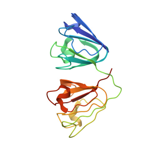
    We describe a 5-year-old boy with a unique congenital cataract caused by deposition of numerous birefringent, pleiochroic and macroscopically prismatic crystals. Crystal analysis with subsequent automatic Edman degradation and matrix-associated laser desorption ionization time-of-flight mass spectrometry have identified the crystal-forming protein as gammaD-crystallin (CRYGD) lacking the N-terminal methionine. Sequencing of the CRYGD gene has shown a heterozygous C-->A transversion in position 109 of the inferred cDNA (36R-->S transversion of the processed, N-terminal methionine-lacking CRYGD). The lens protein crystals were X-ray diffracting, and our crystal structure solution at 2.25 A suggests that mutant R36S CRYGD has an unaltered protein fold. In contrast, the observed crystal packing is possible only with the mutant protein molecules that lack the bulky Arg36 side chain. This is the first described case of human cataract caused by crystallization of a protein in the lens. It involves the third known mutation in the CRYGD gene but offers, for the first time, a causative explanation of the phenotype.


  • Organizational Affiliation

    Institute of Inherited Metabolic Diseases, Division B, Building D, Ke Karlovu 2, 12808 Prague 2, Czech Republic.


Macromolecules
Find similar proteins by:  (by identity cutoff)  |  3D Structure
Entity ID: 1
MoleculeChains Sequence LengthOrganismDetailsImage
Gamma crystallin D
A, B
173Homo sapiensMutation(s): 1 
UniProt & NIH Common Fund Data Resources
Find proteins for P07320 (Homo sapiens)
Explore P07320 
Go to UniProtKB:  P07320
PHAROS:  P07320
GTEx:  ENSG00000118231 
Entity Groups  
Sequence Clusters30% Identity50% Identity70% Identity90% Identity95% Identity100% Identity
UniProt GroupP07320
Sequence Annotations
Expand
  • Reference Sequence
Experimental Data & Validation

Experimental Data

  • Method: X-RAY DIFFRACTION
  • Resolution: 2.20 Å
  • R-Value Free: 0.257 
  • R-Value Work: 0.197 
  • R-Value Observed: 0.200 
  • Space Group: P 21 21 21
Unit Cell:
Length ( Å )Angle ( ˚ )
a = 54.375α = 90
b = 81.778β = 90
c = 106.279γ = 90
Software Package:
Software NamePurpose
REFMACrefinement
DENZOdata reduction
SCALEPACKdata scaling
EPMRphasing

Structure Validation

View Full Validation Report


Entry History 

Deposition Data

Revision History  (Full details and data files)

  • Version 1.0: 2006-05-02
    Type: Initial release
  • Version 1.1: 2008-05-01
    Changes: Version format compliance
  • Version 1.2: 2011-07-13
    Changes: Advisory, Version format compliance
  • Version 1.3: 2021-10-20
    Changes: Database references
  • Version 1.4: 2023-08-30
    Changes: Data collection, Refinement description