2FLN

binary complex of catalytic core of human DNA polymerase iota with DNA (template A)


Experimental Data Snapshot

  • Method: X-RAY DIFFRACTION
  • Resolution: 2.50 Å
  • R-Value Free: 0.279 
  • R-Value Work: 0.226 
  • R-Value Observed: 0.231 

wwPDB Validation   3D Report Full Report


This is version 1.3 of the entry. See complete history


Literature

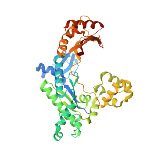
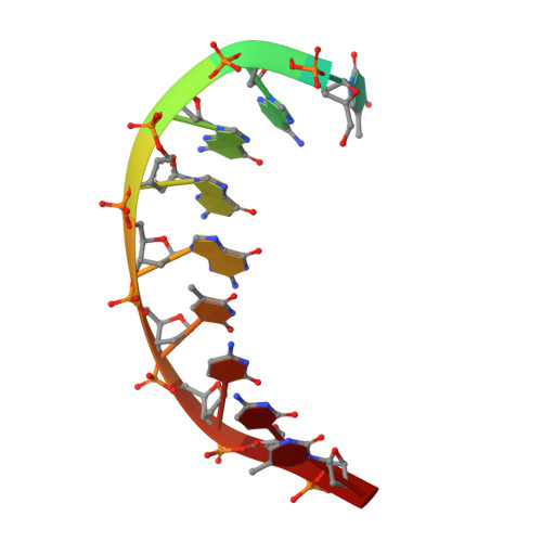
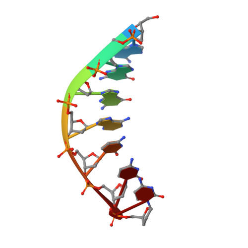
An incoming nucleotide imposes an anti to syn conformational change on the templating purine in the human DNA polymerase-iota active site.

Nair, D.T.Johnson, R.E.Prakash, L.Prakash, S.Aggarwal, A.K.

(2006) Structure 14: 749-755

  • DOI: https://doi.org/10.1016/j.str.2006.01.010
  • Primary Citation of Related Structures:  
    2FLL, 2FLN, 2FLP

  • PubMed Abstract: 

    Substrate-induced conformational change of the protein is the linchpin of enzymatic reactions. Replicative DNA polymerases, for example, convert from an open to a closed conformation in response to dNTP binding. Human DNA polymerase-iota (hPoliota), a member of the Y family of DNA polymerases, differs strikingly from other polymerases in its much higher proficiency and fidelity for nucleotide incorporation opposite template purines than opposite template pyrimidines. We present here a crystallographic analysis of hPoliota binary complexes, which together with the ternary complexes show that, contrary to replicative DNA polymerases, the DNA, and not the polymerase, undergoes the primary substrate-induced conformational change. The incoming dNTP "pushes" templates A and G from the anti to the syn conformation dictated by a rigid hPoliota active site. Together, the structures posit a mechanism for template selection wherein dNTP binding induces a conformational switch in template purines for productive Hoogsteen base pairing.


  • Organizational Affiliation

    Structural Biology Program, Department of Physiology and Biophysics, Mount Sinai School of Medicine, Box 1677, 1425 Madison Avenue, New York, New York 10029, USA.


Macromolecules

Find similar proteins by:  (by identity cutoff)  |  3D Structure
Entity ID: 3
MoleculeChains Sequence LengthOrganismDetailsImage
DNA polymerase iotaC [auth A]420Homo sapiensMutation(s): 0 
Gene Names: POLIRAD30B
EC: 2.7.7.7
UniProt & NIH Common Fund Data Resources
Find proteins for Q9UNA4 (Homo sapiens)
Explore Q9UNA4 
Go to UniProtKB:  Q9UNA4
PHAROS:  Q9UNA4
GTEx:  ENSG00000101751 
Entity Groups  
Sequence Clusters30% Identity50% Identity70% Identity90% Identity95% Identity100% Identity
UniProt GroupQ9UNA4
Sequence Annotations
Expand
  • Reference Sequence

Find similar nucleic acids by:  Sequence   |   3D Structure  

Entity ID: 1
MoleculeChains LengthOrganismImage
DNA template strandA [auth T]11N/A
Sequence Annotations
Expand
  • Reference Sequence

Find similar nucleic acids by:  Sequence   |   3D Structure  

Entity ID: 2
MoleculeChains LengthOrganismImage
DNA primer strandB [auth P]7N/A
Sequence Annotations
Expand
  • Reference Sequence
Experimental Data & Validation

Experimental Data

  • Method: X-RAY DIFFRACTION
  • Resolution: 2.50 Å
  • R-Value Free: 0.279 
  • R-Value Work: 0.226 
  • R-Value Observed: 0.231 
  • Space Group: P 65 2 2
Unit Cell:
Length ( Å )Angle ( ˚ )
a = 98.114α = 90
b = 98.114β = 90
c = 202.823γ = 120
Software Package:
Software NamePurpose
REFMACrefinement
HKL-2000data reduction
SCALEPACKdata scaling
AMoREphasing

Structure Validation

View Full Validation Report


Entry History 

Deposition Data

Revision History  (Full details and data files)

  • Version 1.0: 2006-12-05
    Type: Initial release
  • Version 1.1: 2008-05-01
    Changes: Version format compliance
  • Version 1.2: 2011-07-13
    Changes: Advisory, Version format compliance
  • Version 1.3: 2023-08-30
    Changes: Data collection, Database references, Derived calculations, Refinement description