Calcium ion exchange in crystalline gelsolin
Chumnarnsilpa, S., Loonchanta, A., Xue, B., Choe, H., Urosev, D., Wang, H., Lindberg, U., Burtnick, L.D., Robinson, R.C.(2006) J Mol Biol 357: 773-782
- PubMed: 16466744
- DOI: https://doi.org/10.1016/j.jmb.2006.01.026
- Primary Citation of Related Structures:
2FH1, 2FH2, 2FH3, 2FH4 - PubMed Abstract:
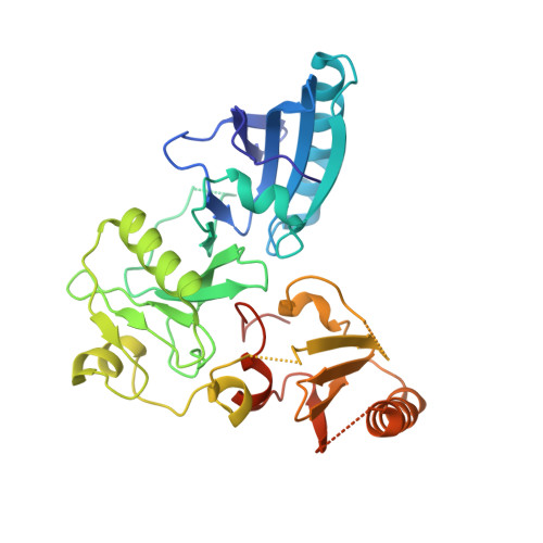
Gelsolin is a calcium and pH-sensitive modulator of actin filament length. Here, we use X-ray crystallography to examine the extraction and exchange of calcium ions from their binding sites in different crystalline forms of the activated N and C-terminal halves of gelsolin, G1-G3 and G4-G6, respectively. We demonstrate that the combination of calcium and low pH activating conditions do not induce conformational changes in G4-G6 beyond those elicited by calcium alone. EGTA is able to remove calcium ions bound to the type I and type II metal ion-binding sites in G4-G6. Constrained by crystal contacts and stabilized by interdomain interaction surfaces, the gross structure of calcium-depleted G4-G6 remains that of the activated form. However, high-resolution details of changes in the ion-binding sites may represent the initial steps toward restoration of the arrangement of domains found in the calcium-free inactive form of gelsolin in solution. Furthermore, bathing crystals with the trivalent calcium ion mimic, Tb3+, results in anomalous scattering data that permit unequivocal localization of terbium ions in each of the proposed type I and type II ion-binding sites of both halves of gelsolin. In contrast to predictions based on solution studies, we find that no calcium ion is immune to exchange.
Organizational Affiliation:
Department of Medical Biochemistry and Microbiology, Uppsala Biomedical Center, Uppsala University, Uppsala 751 23, Sweden.