The sec14 homology module of neurofibromin binds cellular glycerophospholipids: mass spectrometry and structure of a lipid complex
Welti, S., Fraterman, S., D'Angelo, I., Wilm, M., Scheffzek, K.(2007) J Mol Biol 366: 551-562
- PubMed: 17187824
- DOI: https://doi.org/10.1016/j.jmb.2006.11.055
- Primary Citation of Related Structures:
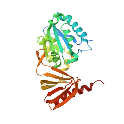
2E2X - PubMed Abstract:
Neurofibromin is the protein product of the tumor suppressor gene NF1, alterations of which are responsible for the pathogenesis of the common disorder Neurofibromatosis type I (NF1). The only well-characterized function of neurofibromin is its RasGAP activity, contained in the central GAP related domain (GRD). By solving the crystal structure of a 31 kDa fragment at the C-terminal end of the GRD we have recently identified a novel bipartite lipid-binding module composed of a Sec14 homologous and a previously undetected pleckstrin homology (PH)-like domain. Using lipid exchange assays along with mass spectrometry we show here that the Sec14-like portion binds to 1-(3-sn-phosphatidyl)-sn-glycerol (PtdGro), (3-sn-phosphatidyl)-ethanolamine (PtdEtn) and -choline (PtdCho) and to a minor extent to (3-sn-phosphatidyl)-l-serine (PtdSer) and 1-(3-sn-phosphatidyl)-d-myo-inositol (PtdIns). Phosphorylated PtdIns (PtdInsPs) are not detected as binders in the mass spectrometry assay, but their soluble inositol-phosphate headgroups and related compounds can inhibit the lipid exchange reaction. We also present here the crystal structure of this module with the Sec14 portion bound to a cellular glycerophospholipid ligand. Our structure has model character for the substrate-bound form of yeast Sec14p, of which only detergent bound structures are available so far. To assess potential regulation of the lipid exchange reaction in detail, we present a novel strategy using nanospray mass spectrometry. Ion intensities of initial phospholipids and exchanged deuterated analogues bound by the protein module allow the quantitative analysis of differences in the exchange activity under various conditions.
Organizational Affiliation:
Structural and Computational Biology, Developmental Biology and Gene Expression Units, European Molecular Biology Laboratory, Meyerhofstrasse 1, 69117 Heidelberg, Germany.