Solution structure of a band 3 peptide inhibitor bound to aldolase: a proposed mechanism for regulating binding by tyrosine phosphorylation.
Schneider, M.L., Post, C.B.(1995) Biochemistry 34: 16574-16584
- PubMed: 8527430
- DOI: https://doi.org/10.1021/bi00051a005
- Primary Citation of Related Structures:
2BTA, 2BTB - PubMed Abstract:
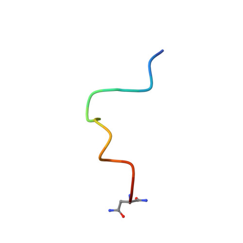
Human erythrocyte band 3 inhibits glycolytic enzymes, including aldolase, by binding these cytoplasmic enzymes at its N-terminus. Phosphorylation of Y8 disrupts inhibition, and there is evidence that in vivo glycolysis levels in erythrocytes are regulated in part by a phosphorylation/dephosphorylation signaling pathway. The structural basis for control by phosphorylation has been investigated by NMR studies on a complex between aldolase and a synthetic peptide corresponding to the first 15 residues of band 3 (MEELQDDYEDMMEEN-NH2). The structure of this band 3 peptide (B3P) when it is bound to rabbit muscle aldolase was determined using the exchange-transferred nuclear Overhauser effect (ETNOE). Two hundred NMR structures for B3P were generated by simulated annealing molecular dynamics with NMR-derived distance restraints and excluding electrostatic terms. Twenty structures were further refined against a force field including full partial charges. The important conformational feature of B3P in the bound state is a folded loop structure involving residues 4-9 and M12 that surrounds Y8 and is stabilized by a hydrophobic cluster with the ring of Y8 sandwiched between the methyl groups of L4 and M12. Differential line broadening indicates that this loop structure binds aldolase in a relatively specific manner, while terminal regions are structurally heterogeneous. To better understand B3P inhibition of aldolase and the mechanism of phosphorylation control, a complex was modeled by docking B3P into the active site of aldolase and optimizing the fit using restrained molecular dynamics and energy minimization. The B3P loop is complementary in conformation to the beta-barrel central core containing the aldolase active site residues. Binding is electrostatic in nature with numerous ionic and hydrogen-bonding interactions involving several conserved lysine and arginine residues of aldolase. How phosphorylation of band 3 could disrupt inhibition was considered by modeling a phosphoryl moiety onto Y8 of B3P. An energetic analysis with respect to rigid phosphate rotation suggests that aldolase inhibition is reversed primarily because of electrostatic repulsion between B3P residues that destabilizes the B3P loop formed in the complex. This proposed intramolecular mechanism for blocking protein--protein association by electrostatic repulsion with the phosphoryl group may be applicable to other protein--protein signaling complexes.
Organizational Affiliation:
Department of Medicinal Chemistry, Purdue University, West Lafayette, Indiana 47907-1333, USA.