2AUD

Unliganded HincII


Experimental Data Snapshot

  • Method: X-RAY DIFFRACTION
  • Resolution: 2.10 Å
  • R-Value Free: 0.327 
  • R-Value Work: 0.248 
  • R-Value Observed: 0.248 

wwPDB Validation   3D Report Full Report


This is version 1.5 of the entry. See complete history


Literature

DNA-induced conformational changes in Type II restriction endonucleases: The structure of unliganded HincII

Horton, N.C.Little, E.J.

(2005) J Mol Biol 351: 76-88

  • DOI: https://doi.org/10.1016/j.jmb.2005.05.063
  • Primary Citation of Related Structures:  
    2AUD

  • PubMed Abstract: 

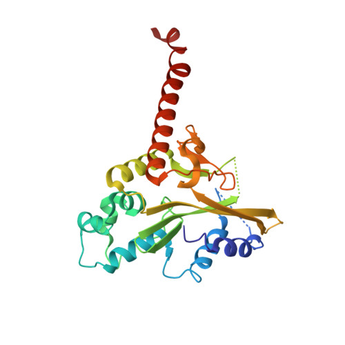
    The 2.1A crystal structure of the unliganded type II restriction endonuclease, HincII, is described. Although the asymmetric unit contains only a single monomer, crystal lattice contacts bring two monomers together to form a dimer very similar to that found in the DNA bound form. Comparison with the published DNA bound structure reveals that the DNA binding pocket is expanded in the unliganded structure. Comparison of the unliganded and DNA liganded structures reveals a simple rotation of subunits by 11 degrees each, or 22 degrees total, to a more closed structure around the bound DNA. Comparison of this conformational change to that observed in the other type II restriction endonucleases where DNA bound and unliganded forms have both been characterized, shows considerable variation in the types of conformational changes that can occur. The conformational changes in three can be described by a simple rotation of subunits, while in two others both rotation and translation of subunits relative to one another occurs. In addition, the endonucleases having subunits that undergo the greatest amount of rotation upon DNA binding are found to be those that distort the bound DNA the least, suggesting that DNA bending may be less facile in dimers possessing greater flexibility.


  • Organizational Affiliation

    Department of Biochemistry and Molecular Biophysics, University of Arizona, Tucson, AZ 85721, USA.


Macromolecules
Find similar proteins by:  (by identity cutoff)  |  3D Structure
Entity ID: 1
MoleculeChains Sequence LengthOrganismDetailsImage
Type II restriction enzyme HincII257Haemophilus influenzaeMutation(s): 0 
Gene Names: hincIIR
EC: 3.1.21.4
UniProt
Find proteins for P17743 (Haemophilus influenzae)
Explore P17743 
Go to UniProtKB:  P17743
Entity Groups  
Sequence Clusters30% Identity50% Identity70% Identity90% Identity95% Identity100% Identity
UniProt GroupP17743
Sequence Annotations
Expand
  • Reference Sequence
Experimental Data & Validation

Experimental Data

  • Method: X-RAY DIFFRACTION
  • Resolution: 2.10 Å
  • R-Value Free: 0.327 
  • R-Value Work: 0.248 
  • R-Value Observed: 0.248 
  • Space Group: P 43 21 2
Unit Cell:
Length ( Å )Angle ( ˚ )
a = 59.74α = 90
b = 59.74β = 90
c = 164.44γ = 90
Software Package:
Software NamePurpose
MAR345data collection
SCALEPACKdata scaling
CNSrefinement
CNSphasing

Structure Validation

View Full Validation Report


Entry History 

Deposition Data

Revision History  (Full details and data files)

  • Version 1.0: 2005-10-04
    Type: Initial release
  • Version 1.1: 2008-05-01
    Changes: Version format compliance
  • Version 1.2: 2011-07-13
    Changes: Derived calculations, Version format compliance
  • Version 1.3: 2011-11-16
    Changes: Atomic model
  • Version 1.4: 2017-10-11
    Changes: Refinement description
  • Version 1.5: 2023-08-23
    Changes: Data collection, Database references, Refinement description