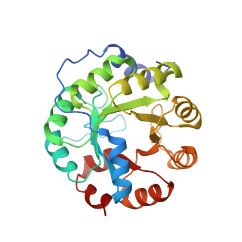
The catalytic mechanism of indole-3-glycerol phosphate synthase: Crystal structures of complexes of the enzyme from Sulfolobus solfataricus with the substrate analogue, substrate, and product
Hennig, M., Darimont, B.D., Jansonius, J.N., Kirschner, K.(2002) J Mol Biol 319: 757-766
- PubMed: 12054868
- DOI: https://doi.org/10.1016/S0022-2836(02)00378-9
- Primary Citation of Related Structures:
1A53, 1LBF, 1LBL - PubMed Abstract:
Indoleglycerol phosphate synthase catalyzes the ring closure of an N-alkylated anthranilate to a 3-alkyl indole derivative, a reaction requiring Lewis acid catalysis in vitro. Here, we investigated the enzymatic reaction mechanism through X-ray crystallography of complexes of the hyperthermostable enzyme from Sulfolobus solfataricus with the substrate 1-(o-carboxyphenylamino) 1-deoxyribulose 5-phosphate, a substrate analogue and the product indole-3-glycerol phosphate. The substrate and the substrate analogue are bound to the active site in a similar, extended conformation between the previously identified phosphate binding site and a hydrophobic pocket for the anthranilate moiety. This binding mode is unproductive, because the carbon atoms that are to be joined are too far apart. The indole ring of the bound product resides in a second hydrophobic pocket adjacent to that of the anthranilate moiety of the substrate. Although the hydrophobic moiety of the substrate moves during catalysis from one hydrophobic pocket to the other, the triosephosphate moiety remains rigidly bound to the same set of hydrogen-bonding residues. Simultaneously, the catalytically important residues Lys53, Lys110 and Glu159 maintain favourable distances to the atoms of the ligand undergoing covalent changes. On the basis of these data, the structures of two putative catalytic intermediates were modelled into the active site. This new structural information and the modelling studies provide further insight into the mechanism of enzyme-catalyzed indole synthesis. The charged epsilon-amino group of Lys110 is the general acid, and the carboxylate group of Glu159 is the general base. Lys53 guides the substrate undergoing conformational transitions during catalysis, by forming a salt-bridge to the carboxylate group of its anthranilate moiety.
Organizational Affiliation:
Division Structural Biology, Biozentrum, University of Basel, Klingelbergstrasse 70, CH-4056 Basel, Switzerland. michael.hennig@roche.com