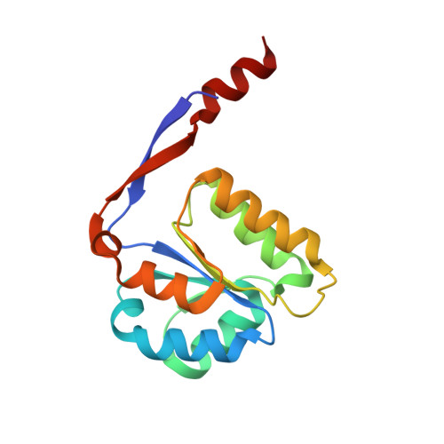
Mechanistic implications of methylglyoxal synthase complexed with phosphoglycolohydroxamic acid as observed by X-ray crystallography and NMR spectroscopy.
Marks, G.T., Harris, T.K., Massiah, M.A., Mildvan, A.S., Harrison, D.H.(2001) Biochemistry 40: 6805-6818
- PubMed: 11389594
- DOI: https://doi.org/10.1021/bi0028237
- Primary Citation of Related Structures:
1IK4 - PubMed Abstract:
Methylglyoxal synthase (MGS) and triosephosphate isomerase (TIM) share neither sequence nor structural similarities, yet the reactions catalyzed by both enzymes are similar, in that both initially convert dihydroxyacetone phosphate to a cis-enediolic intermediate. This enediolic intermediate is formed from the abstraction of the pro-S C3 proton of DHAP by Asp-71 of MGS or the pro-R C3 proton of DHAP by Glu-165 of TIM. MGS then catalyzes the elimination of phosphate from this enediolic intermediate to form the enol of methylglyoxal, while TIM catalyzes proton donation to C2 to form D-glyceraldehyde phosphate. A competitive inhibitor of TIM, phosphoglycolohydroxamic acid (PGH) is found to be a tight binding competitive inhibitor of MGS with a K(i) of 39 nM. PGH's high affinity for MGS may be due in part to a short, strong hydrogen bond (SSHB) from the NOH of PGH to the carboxylate of Asp-71. Evidence for this SSHB is found in X-ray, 1H NMR, and fractionation factor data. The X-ray structure of the MGS homohexamer complexed with PGH at 2.0 A resolution shows this distance to be 2.30-2.37 +/- 0.24 A. 1H NMR shows a PGH-dependent 18.1 ppm signal that is consistent with a hydrogen bond length of 2.49 +/- 0.02 A. The D/H fractionation factor (phi = 0.43 +/- 0.02) is consistent with a hydrogen bond length of 2.53 +/- 0.01 A. Further, 15N NMR suggests a significant partial positive charge on the nitrogen atom of bound PGH, which could strengthen hydrogen bond donation to Asp-71. Both His-98 and His-19 are uncharged in the MGS-PGH complex on the basis of the chemical shifts of their Cdelta and C(epsilon) protons. The crystal structure reveals that Asp-71, on the re face of PGH, and His-19, on the si face of PGH, both approach the NO group of the analogue, while His-98, in the plane of PGH, approaches the carbonyl oxygen of the analogue. The phosphate group of PGH accepts nine hydrogen bonds from seven residues and is tilted out of the imidate plane of PGH toward the re face. Asp-71 and phosphate are thus positioned to function as the base and leaving group, respectively, in a concerted suprafacial 1,4-elimination of phosphate from the enediolic intermediate in the second step of the MGS reaction. Combined, these data suggest that Asp-71 is the one base that initially abstracts the C3 pro-S proton from DHAP and subsequently the 3-OH proton from the enediolic intermediate. This mechanism is compared to an alternative TIM-like mechanism for MGS, and the relative merits of both mechanisms are discussed.
Organizational Affiliation:
Department of Biochemistry, Medical College of Wisconsin, 8701 Watertown Plank Road, Milwaukee, Wisconsin 53226-3548, USA.