1GZD

Crystal structure of pig phosphoglucose isomerase


Experimental Data Snapshot

  • Method: X-RAY DIFFRACTION
  • Resolution: 2.50 Å
  • R-Value Free: 0.241 
  • R-Value Work: 0.167 
  • R-Value Observed: 0.175 

wwPDB Validation   3D Report Full Report


This is version 1.3 of the entry. See complete history


Literature

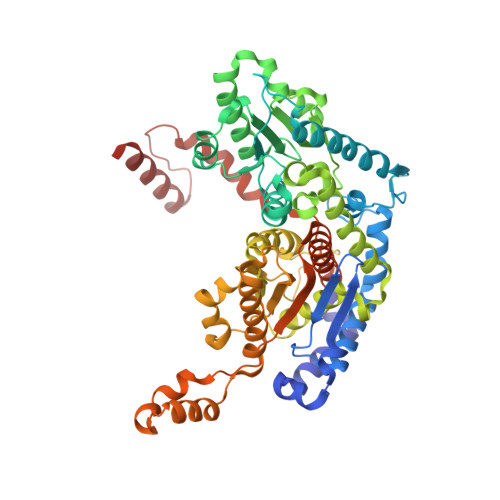
Crystal Structure of Phosphoglucose Isomerase from Pig Muscle and its Complex with 5-Phosphoarabinonate

Davies, C.Muirhead, H.

(2002) Proteins 49: 577


Macromolecules
Find similar proteins by:  (by identity cutoff)  |  3D Structure
Entity ID: 1
MoleculeChains Sequence LengthOrganismDetailsImage
GLUCOSE-6-PHOSPHATE ISOMERASE557Sus scrofaMutation(s): 0 
EC: 5.3.1.9
UniProt
Find proteins for P08059 (Sus scrofa)
Explore P08059 
Go to UniProtKB:  P08059
Entity Groups  
Sequence Clusters30% Identity50% Identity70% Identity90% Identity95% Identity100% Identity
UniProt GroupP08059
Sequence Annotations
Expand
  • Reference Sequence
Small Molecules
Ligands 1 Unique
IDChains Name / Formula / InChI Key2D Diagram3D Interactions
SO4
Query on SO4

Download Ideal Coordinates CCD File 
B [auth A]SULFATE ION
O4 S
QAOWNCQODCNURD-UHFFFAOYSA-L
Experimental Data & Validation

Experimental Data

  • Method: X-RAY DIFFRACTION
  • Resolution: 2.50 Å
  • R-Value Free: 0.241 
  • R-Value Work: 0.167 
  • R-Value Observed: 0.175 
  • Space Group: P 43 21 2
Unit Cell:
Length ( Å )Angle ( ˚ )
a = 95.2α = 90
b = 95.2β = 90
c = 138.3γ = 90
Software Package:
Software NamePurpose
REFMACrefinement
MOSFLMdata reduction
SCALAdata scaling

Structure Validation

View Full Validation Report


Entry History 

Deposition Data

Revision History  (Full details and data files)

  • Version 1.0: 2002-06-10
    Type: Initial release
  • Version 1.1: 2011-12-28
    Changes: Database references, Derived calculations, Non-polymer description, Other, Version format compliance
  • Version 1.2: 2014-02-19
    Changes: Database references
  • Version 1.3: 2019-05-15
    Changes: Data collection, Experimental preparation, Other