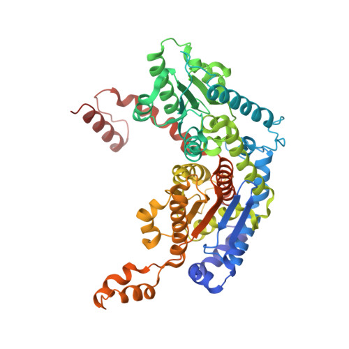
1G98

CRYSTAL STRUCTURE ANALYSIS OF RABBIT PHOSPHOGLUCOSE ISOMERASE COMPLEXED WITH 5-PHOSPHOARABINONATE, A TRANSITION STATE ANALOGUE


Experimental Data Snapshot

  • Method: X-RAY DIFFRACTION
  • Resolution: 1.90 Å
  • R-Value Free: 0.236 
  • R-Value Work: 0.211 

wwPDB Validation   3D Report Full Report


This is version 1.4 of the entry. See complete history


Literature

Crystal structure of rabbit phosphoglucose isomerase complexed with 5-phospho-D-arabinonate identifies the role of Glu357 in catalysis.

Jeffery, C.J.Hardre, R.Salmon, L.

(2001) Biochemistry 40: 1560-1566

  • DOI: https://doi.org/10.1021/bi0018483
  • Primary Citation of Related Structures:  
    1G98

  • PubMed Abstract: 

    Phosphoglucose isomerase (PGI; E.C. 5.3.1.9) catalyzes the second step in glycolysis, the interconversion of D-glucose-6-phosphate and D-fructose-6-phosphate. We determined the X-ray crystal structure of rabbit PGI complexed with a competitive inhibitor of isomerase activity, 5-phospho-D-arabinonate (5PAA), at 1.9 A resolution. 5PAA is a better mimic of the proposed cis-enediol(ate) intermediate than 6-phospho-D-gluconate, which was used in a previously reported crystal structure of rabbit PGI. The orientation of 5PAA bound in the enzyme active site predicts that active site residue Glu357 is the residue that transfers a proton between C2 and C1 of the proposed cis-enediol(ate) intermediate. Amino acid residues Arg272 and Lys210 are predicted to be involved in stabilizing the negative charge of the intermediate.


  • Organizational Affiliation

    Laboratory for Molecular Biology, MC567, Department of Biology, University of Illinois at Chicago, Chicago, IL 60607, USA. cjeffery@uic.edu


Macromolecules
Find similar proteins by:  (by identity cutoff)  |  3D Structure
Entity ID: 1
MoleculeChains Sequence LengthOrganismDetailsImage
PHOSPHOGLUCOSE ISOMERASE
A, B
558Oryctolagus cuniculusMutation(s): 0 
EC: 5.3.1.9
UniProt
Find proteins for Q9N1E2 (Oryctolagus cuniculus)
Explore Q9N1E2 
Go to UniProtKB:  Q9N1E2
Entity Groups  
Sequence Clusters30% Identity50% Identity70% Identity90% Identity95% Identity100% Identity
UniProt GroupQ9N1E2
Sequence Annotations
Expand
  • Reference Sequence
Small Molecules
Ligands 1 Unique
IDChains Name / Formula / InChI Key2D Diagram3D Interactions
PA5
Query on PA5

Download Ideal Coordinates CCD File 
C [auth A],
D [auth B]
5-PHOSPHOARABINONIC ACID
C5 H11 O9 P
HNECGPFIYSOYHF-JJYYJPOSSA-N
Binding Affinity Annotations 
IDSourceBinding Affinity
PA5 Binding MOAD:  1G98 Ki: 2000 (nM) from 1 assay(s)
PDBBind:  1G98 Ki: 2000 (nM) from 1 assay(s)
Experimental Data & Validation

Experimental Data

  • Method: X-RAY DIFFRACTION
  • Resolution: 1.90 Å
  • R-Value Free: 0.236 
  • R-Value Work: 0.211 
  • Space Group: C 2 2 21
Unit Cell:
Length ( Å )Angle ( ˚ )
a = 82.829α = 90
b = 116.554β = 90
c = 271.824γ = 90
Software Package:
Software NamePurpose
CNSrefinement
DENZOdata reduction
SCALEPACKdata scaling
CNSphasing

Structure Validation

View Full Validation Report


Entry History 

Deposition Data

Revision History  (Full details and data files)

  • Version 1.0: 2001-06-01
    Type: Initial release
  • Version 1.1: 2008-04-27
    Changes: Version format compliance
  • Version 1.2: 2011-07-13
    Changes: Version format compliance
  • Version 1.3: 2011-11-16
    Changes: Atomic model
  • Version 1.4: 2023-08-09
    Changes: Data collection, Database references, Derived calculations, Refinement description