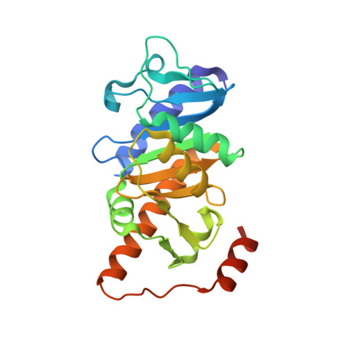
Structures of l-fuculose-1-phosphate aldolase mutants outlining motions during catalysis.
Joerger, A.C., Mueller-Dieckmann, C., Schulz, G.E.(2000) J Mol Biol 303: 531-543
- PubMed: 11054289
- DOI: https://doi.org/10.1006/jmbi.2000.4153
- Primary Citation of Related Structures:
1E46, 1E47, 1E48, 1E49, 1E4A, 1E4B, 1E4C - PubMed Abstract:
The crystal structures of l-fuculose-1-phosphate aldolase (FucA) with and without a ligated analogue of dihydroxyacetone phosphate (DHAP) and of a number of active center mutants have resulted in a model of the catalytic mechanism. This model has now been confirmed by structural analyses of further mutations at the zinc coordination sphere and at the phosphate site. In addition, these mutants have revealed new aspects of the catalysis: the hydroxyl group of Tyr113' (from a neighboring subunit), which sits just outside the zinc coordination sphere, steers DHAP towards a productive binding mode at the zinc ion; Glu73 contacts zinc in between the two ligand positions intended for the DHAP oxygen atoms and thus avoids blocking of these positions by a tetrahedrally coordinated hydroxy ion; the FucA polypeptide does not assume its minimum energy state but oscillates between two states of elevated energy as demonstrated by a mutant in a minimum energy state. The back and forth motion involves a mobile loop connecting the phosphate site with intersubunit motions and thus with the Brownian motion of the solvent. The phosphate group is bound strongly at a given distance to the zinc ion, which prevents the formation of too tight a DHAP:zinc complex. This observation explains our failure to find mutants that accept phosphate-free substitutes for DHAP. The FucA zinc coordination sphere is compared with that of carbonic anhydrase.
Organizational Affiliation:
Institut für Organische Chemie und Biochemie, Freiburg im Breisgau, Germany-79104, USA.