1D6S

CRYSTAL STRUCTURE OF THE K41A MUTANT OF O-ACETYLSERINE SULFHYDRYLASE COMPLEXED IN EXTERNAL ALDIMINE LINKAGE WITH METHIONINE


Experimental Data Snapshot

  • Method: X-RAY DIFFRACTION
  • Resolution: 2.30 Å
  • R-Value Free: 0.223 
  • R-Value Work: 0.174 
  • R-Value Observed: 0.174 

wwPDB Validation   3D Report Full Report


Ligand Structure Quality Assessment 


This is version 1.4 of the entry. See complete history


Literature

Ligand binding induces a large conformational change in O-acetylserine sulfhydrylase from Salmonella typhimurium.

Burkhard, P.Tai, C.H.Ristroph, C.M.Cook, P.F.Jansonius, J.N.

(1999) J Mol Biol 291: 941-953

  • DOI: https://doi.org/10.1006/jmbi.1999.3002
  • Primary Citation of Related Structures:  
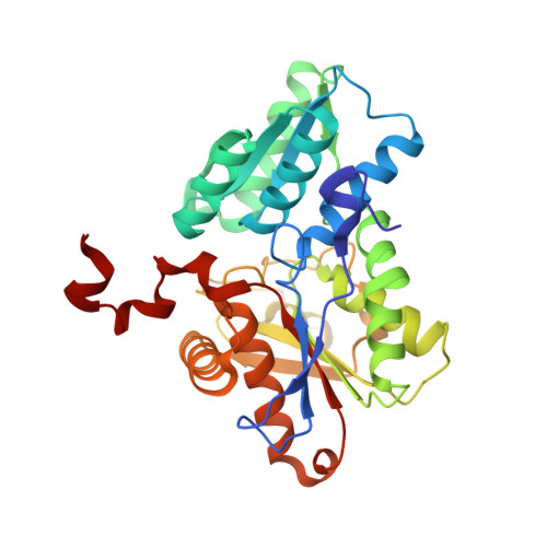
    1D6S

  • PubMed Abstract: 

    Covalent binding of L-methionine as an external aldimine to the pyridoxal 5'-phosphate-cofactor in the K41A mutant of O-acetylserine sulfhydrylase from Salmonella typhimurium induces a large conformational change in the protein. Methionine mimics the action of the substrate O-acetyl-L-serine during catalysis. The alpha-carboxylate moiety of L-methionine in external aldimine linkage with the active site pyridoxal 5'-phosphate forms a hydrogen bonding network to the "asparagine-loop" P67-T68-N69-G70 which adopts a different conformation than in the native protein. The side-chain nitrogen of Asn69 moves more than 7 A to make a hydrogen bond to the alpha-carboxylate group of the inhibitor. As the external aldimine is formed, the PLP tilts by 13 degrees along its longitudinal axis such that C4' moves toward the entrance to the active site and the side-chain of the methionine is directed toward the active site entrance. The local rearrangement acts as a trigger to induce a large global conformational change in the protein. A subdomain comprised of beta-strand 4, alpha-helix 3, beta-strand 5 and alpha-helix 4 moves towards the active site by a rotation of 7 degrees. This subdomain movement results in a reduction of the severe twist of its central beta-sheet and reduces the active site entrance to a small hole, giving access only to small molecules like sulfide, the second substrate, or acetate, the first product.


  • Organizational Affiliation

    Biozentrum, University of Basel, Klingelbergstrasse 70, Basel, CH-4056, Switzerland.


Macromolecules
Find similar proteins by:  (by identity cutoff)  |  3D Structure
Entity ID: 1
MoleculeChains Sequence LengthOrganismDetailsImage
O-ACETYLSERINE SULFHYDRYLASE
A, B
322Salmonella enterica subsp. enterica serovar TyphimuriumMutation(s): 1 
EC: 4.2.99.8
UniProt
Find proteins for P0A1E3 (Salmonella typhimurium (strain LT2 / SGSC1412 / ATCC 700720))
Explore P0A1E3 
Go to UniProtKB:  P0A1E3
Entity Groups  
Sequence Clusters30% Identity50% Identity70% Identity90% Identity95% Identity100% Identity
UniProt GroupP0A1E3
Sequence Annotations
Expand
  • Reference Sequence
Small Molecules
Experimental Data & Validation

Experimental Data

  • Method: X-RAY DIFFRACTION
  • Resolution: 2.30 Å
  • R-Value Free: 0.223 
  • R-Value Work: 0.174 
  • R-Value Observed: 0.174 
  • Space Group: P 21 21 2
Unit Cell:
Length ( Å )Angle ( ˚ )
a = 96.94α = 90
b = 149.93β = 90
c = 53.64γ = 90
Software Package:
Software NamePurpose
AMoREphasing
X-PLORrefinement
MOSFLMdata reduction
CCP4data scaling

Structure Validation

View Full Validation Report


Ligand Structure Quality Assessment 


Entry History 

Deposition Data

Revision History  (Full details and data files)

  • Version 1.0: 2000-04-15
    Type: Initial release
  • Version 1.1: 2008-04-27
    Changes: Version format compliance
  • Version 1.2: 2011-07-13
    Changes: Non-polymer description, Version format compliance
  • Version 1.3: 2021-11-03
    Changes: Database references, Derived calculations
  • Version 1.4: 2024-02-07
    Changes: Data collection