1C4V

SELECTIVE NON ELECTROPHILIC THROMBIN INHIBITORS WITH CYCLOHEXYL MOIETIES.


Experimental Data Snapshot

  • Method: X-RAY DIFFRACTION
  • Resolution: 2.10 Å
  • R-Value Work: 0.167 

wwPDB Validation   3D Report Full Report


This is version 1.5 of the entry. See complete history


Literature

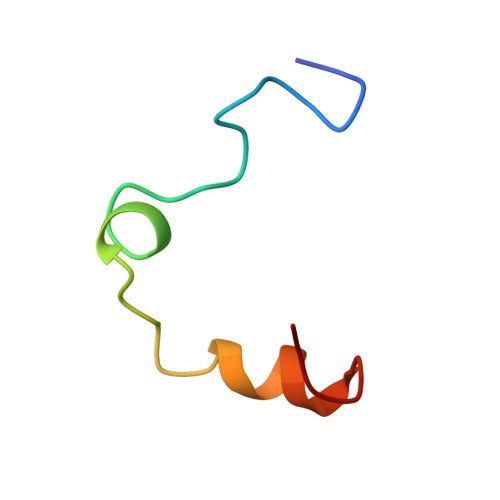
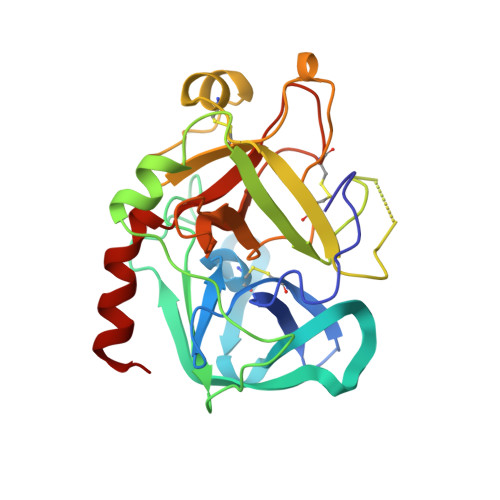
Structure of thrombin complexed with selective non-electrophilic inhibitors having cyclohexyl moieties at P1.

Krishnan, R.Mochalkin, I.Arni, R.Tulinsky, A.

(2000) Acta Crystallogr D Biol Crystallogr 56: 294-303

  • DOI: https://doi.org/10.1107/s0907444900000068
  • Primary Citation of Related Structures:  
    1C4U, 1C4V, 1C4Y, 1D6W, 1D9I

  • PubMed Abstract: 

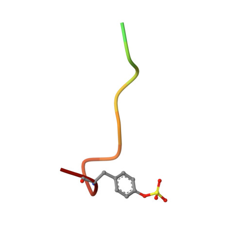
    The crystal structures of five new non-electrophilic beta-strand-templated thrombin active-site inhibitors have been determined bound to the enzyme. Four co-crystallize with hirugen and inhibitor isomorphously to produce thrombin-hirugen crystals (monoclinic, space group C2), while one co-crystallizes in the hexagonal system, space group P6(5). A 1,4-substituted cyclohexyl moiety is conserved at the P1 position of all the inhibitors, along with a fused hetero-bicyclic five- and six-membered ring that occupies the P2 site. Amino, amidino and aminoimidazole groups are attached to the cyclohexyl ring for recognition at the S1 specificity site, while benzylsulfonyl and diphenyl groups enhance the binding at the S3 subsite. The cyclohexyl groups at the P1 positions of three of the inhibitors appear to be in the energetically favored chair conformation, while the imidazole-substituted cyclohexyl rings are in a boat conformation. Somewhat unexpectedly, the two cyclohexyl-aminoimidazole groups bind differently in the specificity site; the unique binding of one is heretofore unreported. The other inhibitors generally mimic arginyl binding at S1. This group of inhibitors combines the non-electrophilicity and selectivity of DAPA-like compounds and the more optimal binding features of the S1-S3 sites of thrombin for peptidic molecules, which results in highly potent (binding constants 12 nM-16 pM, one being 1.1 microM) and selective (ranging from 140 to 20 000 times more selective compared with trypsin) inhibitors of thrombin. The binding modes of these novel inhibitors are correlated with their binding constants, as is their selectivity, in order to provide further insight for the design of therapeutic antithrombotic agents that inhibit thrombin directly at the active site.


  • Organizational Affiliation

    Department of Chemistry, Michigan State University, East Lansing, Michigan 48824-1322, USA.


Macromolecules
Find similar proteins by:  (by identity cutoff)  |  3D Structure
Entity ID: 1
MoleculeChains Sequence LengthOrganismDetailsImage
THROMBIN:SHORT CHAINA [auth 1]36Homo sapiensMutation(s): 0 
EC: 3.4.21.5
UniProt & NIH Common Fund Data Resources
Find proteins for P00734 (Homo sapiens)
Explore P00734 
Go to UniProtKB:  P00734
PHAROS:  P00734
GTEx:  ENSG00000180210 
Entity Groups  
Sequence Clusters30% Identity50% Identity70% Identity90% Identity95% Identity100% Identity
UniProt GroupP00734
Sequence Annotations
Expand
  • Reference Sequence
Find similar proteins by:  (by identity cutoff)  |  3D Structure
Entity ID: 2
MoleculeChains Sequence LengthOrganismDetailsImage
THROMBIN:LONG CHAINB [auth 2]259Homo sapiensMutation(s): 0 
EC: 3.4.21.5
UniProt & NIH Common Fund Data Resources
Find proteins for P00734 (Homo sapiens)
Explore P00734 
Go to UniProtKB:  P00734
PHAROS:  P00734
GTEx:  ENSG00000180210 
Entity Groups  
Sequence Clusters30% Identity50% Identity70% Identity90% Identity95% Identity100% Identity
UniProt GroupP00734
Sequence Annotations
Expand
  • Reference Sequence

Find similar proteins by:  Sequence   |   3D Structure  

Entity ID: 3
MoleculeChains Sequence LengthOrganismDetailsImage
HIRUGENC [auth 3]15N/AMutation(s): 0 
Entity Groups  
Sequence Clusters30% Identity50% Identity70% Identity90% Identity95% Identity100% Identity
Sequence Annotations
Expand
  • Reference Sequence
Small Molecules
Ligands 2 Unique
IDChains Name / Formula / InChI Key2D Diagram3D Interactions
IH2
Query on IH2

Download Ideal Coordinates CCD File 
E [auth 2]2-(2,2-DIPHENYL-ETHYL)-7-METHYL-1,3-DIOXO-2,3,5,8-TETRAHYDRO-1H-[1,2,4]TRIAZOLO [1,2-A]PYRIDAZINE-5-CARBOXYLIC ACID(4-CARBAMIMIDOYL-CYCLOHEXYLMETHYL)-AMIDE
C30 H36 N6 O3
HKVVMQTWGPLJEV-CVJWPJSTSA-N
NA
Query on NA

Download Ideal Coordinates CCD File 
D [auth 2]SODIUM ION
Na
FKNQFGJONOIPTF-UHFFFAOYSA-N
Modified Residues  1 Unique
IDChains TypeFormula2D DiagramParent
TYS
Query on TYS
C [auth 3]L-PEPTIDE LINKINGC9 H11 N O6 STYR
Binding Affinity Annotations 
IDSourceBinding Affinity
IH2 Binding MOAD:  1C4V Ki: 0.02 (nM) from 1 assay(s)
PDBBind:  1C4V Ki: 0.02 (nM) from 1 assay(s)
Experimental Data & Validation

Experimental Data

  • Method: X-RAY DIFFRACTION
  • Resolution: 2.10 Å
  • R-Value Work: 0.167 
  • Space Group: C 1 2 1
Unit Cell:
Length ( Å )Angle ( ˚ )
a = 71.51α = 90
b = 72.02β = 100.8
c = 72.91γ = 90
Software Package:
Software NamePurpose
X-PLORmodel building
PROLSQrefinement
X-PLORphasing

Structure Validation

View Full Validation Report


Entry History 

Deposition Data

Revision History  (Full details and data files)

  • Version 1.0: 2000-09-27
    Type: Initial release
  • Version 1.1: 2007-10-16
    Changes: Version format compliance
  • Version 1.2: 2011-07-13
    Changes: Atomic model, Database references, Derived calculations, Non-polymer description, Structure summary, Version format compliance
  • Version 1.3: 2017-10-04
    Changes: Refinement description
  • Version 1.4: 2023-08-09
    Changes: Data collection, Database references, Derived calculations, Refinement description
  • Version 1.5: 2023-11-15
    Changes: Data collection