1YC0

short form HGFA with first Kunitz domain from HAI-1


Experimental Data Snapshot

  • Method: X-RAY DIFFRACTION
  • Resolution: 2.60 Å
  • R-Value Free: 0.218 
  • R-Value Work: 0.195 
  • R-Value Observed: 0.196 

wwPDB Validation   3D Report Full Report


This is version 1.3 of the entry. See complete history


Literature

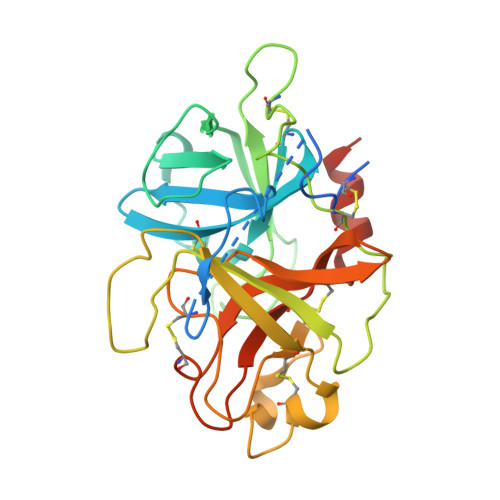
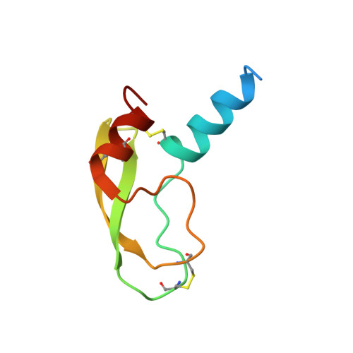
Conformational lability in serine protease active sites: structures of hepatocyte growth factor activator (HGFA) alone and with the inhibitory domain from HGFA inhibitor-1B.

Shia, S.Stamos, J.Kirchhofer, D.Fan, B.Wu, J.Corpuz, R.T.Santell, L.Lazarus, R.A.Eigenbrot, C.

(2005) J Mol Biol 346: 1335-1349

  • DOI: https://doi.org/10.1016/j.jmb.2004.12.048
  • Primary Citation of Related Structures:  
    1YC0

  • PubMed Abstract: 

    Hepatocyte growth factor activator (HGFA) is a serine protease that converts hepatocyte growth factor (HGF) into its active form. When activated HGF binds its cognate receptor Met, cellular signals lead to cell growth, differentiation, and migration, activities which promote tissue regeneration in liver, kidney and skin. Intervention in the conversion of HGF to its active form has the potential to provide therapeutic benefit where HGF/Met activity is associated with tumorigenesis. To help identify ways to moderate HGF/Met effects, we have determined the molecular structure of the protease domain of HGFA. The structure we determined, at 2.7 A resolution, with no pseudo-substrate or inhibitor bound is characterized by an unconventional conformation of key residues in the enzyme active site. In order to find whether this apparently non-enzymatically competent arrangement would persist in the presence of a strongly-interacting inhibitor, we also have determined, at 2.6 A resolution, the X-ray structure of HGFA complexed with the first Kunitz domain (KD1) from the physiological inhibitor hepatocyte growth factor activator inhibitor 1B (HAI-1B). In this complex we observe a rearranged substrate binding cleft that closely mirrors the cleft of other serine proteases, suggesting an extreme conformational dynamism. We also characterize the inhibition of 16 serine proteases by KD1, finding that the previously reported enzyme specificity of the intact extracellular region of HAI-1B resides in KD1 alone. We find that HGFA, matriptase, hepsin, plasma kallikrein and trypsin are potently inhibited, and use the complex structure to rationalize the structural basis of these results.


  • Organizational Affiliation

    Department of Protein Engineering, Genentech, Inc., South San Francisco, CA 94080, USA.


Macromolecules
Find similar proteins by:  (by identity cutoff)  |  3D Structure
Entity ID: 1
MoleculeChains Sequence LengthOrganismDetailsImage
Hepatocyte growth factor activator283Homo sapiensMutation(s): 0 
Gene Names: HGFAC
EC: 3.4.21
UniProt & NIH Common Fund Data Resources
Find proteins for Q04756 (Homo sapiens)
Explore Q04756 
Go to UniProtKB:  Q04756
PHAROS:  Q04756
GTEx:  ENSG00000109758 
Entity Groups  
Sequence Clusters30% Identity50% Identity70% Identity90% Identity95% Identity100% Identity
UniProt GroupQ04756
Sequence Annotations
Expand
  • Reference Sequence
Find similar proteins by:  (by identity cutoff)  |  3D Structure
Entity ID: 2
MoleculeChains Sequence LengthOrganismDetailsImage
Kunitz-type protease inhibitor 1B [auth I]75Homo sapiensMutation(s): 0 
Gene Names: SPINT1HAI1
UniProt & NIH Common Fund Data Resources
Find proteins for O43278 (Homo sapiens)
Explore O43278 
Go to UniProtKB:  O43278
PHAROS:  O43278
GTEx:  ENSG00000166145 
Entity Groups  
Sequence Clusters30% Identity50% Identity70% Identity90% Identity95% Identity100% Identity
UniProt GroupO43278
Sequence Annotations
Expand
  • Reference Sequence
Small Molecules
Ligands 1 Unique
IDChains Name / Formula / InChI Key2D Diagram3D Interactions
PO4
Query on PO4

Download Ideal Coordinates CCD File 
C [auth I]PHOSPHATE ION
O4 P
NBIIXXVUZAFLBC-UHFFFAOYSA-K
Experimental Data & Validation

Experimental Data

  • Method: X-RAY DIFFRACTION
  • Resolution: 2.60 Å
  • R-Value Free: 0.218 
  • R-Value Work: 0.195 
  • R-Value Observed: 0.196 
  • Space Group: P 31 2 1
Unit Cell:
Length ( Å )Angle ( ˚ )
a = 76.218α = 90
b = 76.218β = 90
c = 176.236γ = 120
Software Package:
Software NamePurpose
REFMACrefinement
HKL-2000data reduction
SCALEPACKdata scaling
AMoREphasing

Structure Validation

View Full Validation Report


Entry History 

Deposition Data

Revision History  (Full details and data files)

  • Version 1.0: 2005-02-15
    Type: Initial release
  • Version 1.1: 2008-04-30
    Changes: Version format compliance
  • Version 1.2: 2011-07-13
    Changes: Advisory, Version format compliance
  • Version 1.3: 2023-08-23
    Changes: Data collection, Database references, Derived calculations, Refinement description