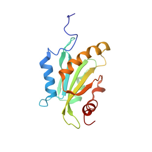
NMR solution structures of actin depolymerizing factor homology domains.
Goroncy, A.K., Koshiba, S., Tochio, N., Tomizawa, T., Sato, M., Inoue, M., Watanabe, S., Hayashizaki, Y., Tanaka, A., Kigawa, T., Yokoyama, S.(2009) Protein Sci 18: 2384-2392
- PubMed: 19768801
- DOI: https://doi.org/10.1002/pro.248
- Primary Citation of Related Structures:
1UDM, 1V6F, 1WFS, 1X67, 2D8B - PubMed Abstract:
Actin is one of the most conserved proteins in nature. Its assembly and disassembly are regulated by many proteins, including the family of actin-depolymerizing factor homology (ADF-H) domains. ADF-H domains can be divided into five classes: ADF/cofilin, glia maturation factor (GMF), coactosin, twinfilin, and Abp1/drebrin. The best-characterized class is ADF/cofilin. The other four classes have drawn much less attention and very few structures have been reported. This study presents the solution NMR structure of the ADF-H domain of human HIP-55-drebrin-like protein, the first published structure of a drebrin-like domain (mammalian), and the first published structure of GMF beta (mouse). We also determined the structures of mouse GMF gamma, the mouse coactosin-like domain and the C-terminal ADF-H domain of mouse twinfilin 1. Although the overall fold of the five domains is similar, some significant differences provide valuable insights into filamentous actin (F-actin) and globular actin (G-actin) binding, including the identification of binding residues on the long central helix. This long helix is stabilized by three or four residues. Notably, the F-actin binding sites of mouse GMF beta and GMF gamma contain two additional beta-strands not seen in other ADF-H structures. The G-actin binding site of the ADF-H domain of human HIP-55-drebrin-like protein is absent and distorted in mouse GMF beta and GMF gamma.
Organizational Affiliation:
RIKEN Systems and Structural Biology Center, 1-7-22 Suehiro-cho, Tsurumi-ku, Yokohama, Kanagawa, Japan.