Molecular insights into the self-assembly mechanism of dystrophia myotonica kinase
Garcia, P., Ucurum, Z., Bucher, R., Svergun, D.I., Huber, T., Lustig, A., Konarev, P.V., Marino, M., Mayans, O.(2006) FASEB J 20: 1142-1151
- PubMed: 16770013
- DOI: https://doi.org/10.1096/fj.05-5262com
- Primary Citation of Related Structures:
1WT6 - PubMed Abstract:
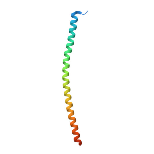
Self-assembly via coiled-coil domains (CC) is crucial for the regulation of the dystrophia myotonica kinase (DMPK) -related family of kinases. These CC domains are thought to form dimeric arrangements and thus to mediate dimerization in these enzymes. Using size exclusion chromatography combined with multiangle static light scattering, we analyzed the oligomeric state of DMPK as well as that of a truncated variant lacking the CC fraction. Remarkably, both forms were found to assemble into robust dimers. In contrast, the CC domain in isolation yielded trimeric assemblies, indicating that the oligomerization properties of CC domains from this kinase family are more diversified than anticipated. The crystal structure of this CC has been elucidated to 1.6 angstroms resolution and its properties in solution established using sedimentation equilibrium and thermal denaturation. These data show that, contrary to expectations, the self-assembly of DMPK is not dictated by the association properties of its CC domain. Instead, it appears to be driven by sequence segments flanking both N and C termini of the catalytic kinase fraction, as suggested by models of head-to-head dimers based on small angle X-ray scattering data. Our findings support a shared pattern of assembly across DMPK, ROCKs, and MRCK members of this family.
Organizational Affiliation:
Division of Structural Biology, Biozentrum, University of Basel, Klingelbergstr. 70, Basel CH-4056, Switzerland.