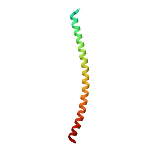
1UII

Crystal structure of Geminin coiled-coil domain


Experimental Data Snapshot

  • Method: X-RAY DIFFRACTION
  • Resolution: 2.00 Å
  • R-Value Free: 0.239 
  • R-Value Work: 0.216 
  • R-Value Observed: 0.216 

wwPDB Validation   3D Report Full Report


This is version 1.3 of the entry. See complete history


Literature

A dimerized coiled-coil domain and an adjoining part of geminin interact with two sites on Cdt1 for replication inhibition

Saxena, S.Yuan, P.Dhar, S.K.Senga, T.Takeda, D.Robinson, H.Kornbluth, S.Swaminathan, K.Dutta, A.

(2004) Mol Cell 15: 245-258

  • DOI: https://doi.org/10.1016/j.molcel.2004.06.045
  • Primary Citation of Related Structures:  
    1UII

  • PubMed Abstract: 

    Geminin is a cellular protein that associates with Cdt1 and inhibits Mcm2-7 loading during S phase. It prevents multiple cycles of replication per cell cycle and prevents episome replication. It also directly inhibits the HoxA11 transcription factor. Here we report that geminin forms a parallel coiled-coil homodimer with atypical residues in the dimer interface. Point mutations that disrupt the dimerization abolish interaction with Cdt1 and inhibition of replication. An array of glutamic acid residues on the coiled-coil domain surface interacts with positive charges in the middle of Cdt1. An adjoining region interacts independently with the N-terminal 100 residues of Cdt1. Both interactions are essential for replication inhibition. The negative residues on the coiled-coil domain and a different part of geminin are also required for interaction with HoxA11. Therefore a rigid cylinder with negative surface charges is a critical component of a bipartite interaction interface between geminin and its cellular targets.


  • Organizational Affiliation

    Department of Biochemistry and Molecular Genetics, University of Virginia, Charlottesville 22908, USA.


Macromolecules
Find similar proteins by:  (by identity cutoff)  |  3D Structure
Entity ID: 1
MoleculeChains Sequence LengthOrganismDetailsImage
Geminin
A, B
83Homo sapiensMutation(s): 0 
Gene Names: GMNN
UniProt & NIH Common Fund Data Resources
Find proteins for O75496 (Homo sapiens)
Explore O75496 
Go to UniProtKB:  O75496
PHAROS:  O75496
GTEx:  ENSG00000112312 
Entity Groups  
Sequence Clusters30% Identity50% Identity70% Identity90% Identity95% Identity100% Identity
UniProt GroupO75496
Sequence Annotations
Expand
  • Reference Sequence
Experimental Data & Validation

Experimental Data

  • Method: X-RAY DIFFRACTION
  • Resolution: 2.00 Å
  • R-Value Free: 0.239 
  • R-Value Work: 0.216 
  • R-Value Observed: 0.216 
  • Space Group: P 21 21 21
Unit Cell:
Length ( Å )Angle ( ˚ )
a = 37.16α = 90
b = 94.46β = 90
c = 102.56γ = 90
Software Package:
Software NamePurpose
DENZOdata reduction
SCALEPACKdata scaling
SOLVEphasing
PHASESphasing
CNSrefinement

Structure Validation

View Full Validation Report


Entry History 

Deposition Data

Revision History  (Full details and data files)

  • Version 1.0: 2004-07-16
    Type: Initial release
  • Version 1.1: 2008-04-27
    Changes: Version format compliance
  • Version 1.2: 2011-07-13
    Changes: Version format compliance
  • Version 1.3: 2023-12-27
    Changes: Data collection, Database references