1TJT

X-ray structure of the human alpha-actinin isoform 3 at 2.2A resolution


Experimental Data Snapshot

  • Method: X-RAY DIFFRACTION
  • Resolution: 2.19 Å
  • R-Value Free: 0.273 
  • R-Value Work: 0.227 
  • R-Value Observed: 0.227 

wwPDB Validation   3D Report Full Report


This is version 1.4 of the entry. See complete history


Literature

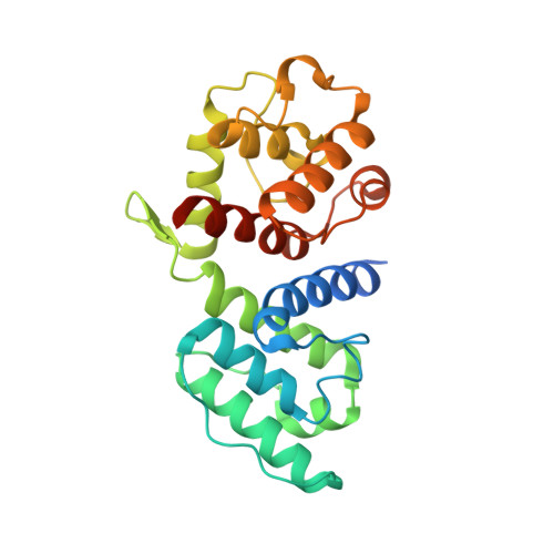
The crystal structure of the actin binding domain from alpha-actinin in its closed conformation: structural insight into phospholipid regulation of alpha-actinin

Franzot, G.Sjoblom, B.Gautel, M.Djinovic Carugo, K.

(2005) J Mol Biol 348: 151-165

  • DOI: https://doi.org/10.1016/j.jmb.2005.01.002
  • Primary Citation of Related Structures:  
    1TJT, 1WKU

  • PubMed Abstract: 

    Alpha-actinin is the major F-actin crosslinking protein in both muscle and non-muscle cells. We report the crystal structure of the actin binding domain of human muscle alpha-actinin-3, which is formed by two consecutive calponin homology domains arranged in a "closed" conformation. Structural studies and available biochemical data on actin binding domains suggest that two calponin homology domains come in a closed conformation in the native apo-form, and that conformational changes involving the relative orientation of the two calponin homology domains are required for efficient binding to actin filaments. The actin binding activity of muscle isoforms is supposed to be regulated by phosphatidylinositol 4,5-bisphosphate (PtdIns(4,5)P2), which binds to the second calponin homology domain. On the basis of structural analysis we propose a distinct binding site for PtdIns(4,5)P2, where the fatty acid moiety would be oriented in a direction that allows it to interact with the linker sequence between the actin binding domain and the first spectrin-like repeat, regulating thereby the binding of the C-terminal calmodulin-like domain to this linker.


  • Organizational Affiliation

    Structural Biology Laboratory, Elettra-Sincrotrone Trieste in Area Science Park, S.S. 14 Km 163,5 34012 Trieste, Italy.


Macromolecules
Find similar proteins by:  (by identity cutoff)  |  3D Structure
Entity ID: 1
MoleculeChains Sequence LengthOrganismDetailsImage
Alpha-actinin 3250Homo sapiensMutation(s): 0 
Gene Names: ACTN3
UniProt & NIH Common Fund Data Resources
Find proteins for Q08043 (Homo sapiens)
Explore Q08043 
Go to UniProtKB:  Q08043
PHAROS:  Q08043
GTEx:  ENSG00000248746 
Entity Groups  
Sequence Clusters30% Identity50% Identity70% Identity90% Identity95% Identity100% Identity
UniProt GroupQ08043
Sequence Annotations
Expand
  • Reference Sequence
Experimental Data & Validation

Experimental Data

  • Method: X-RAY DIFFRACTION
  • Resolution: 2.19 Å
  • R-Value Free: 0.273 
  • R-Value Work: 0.227 
  • R-Value Observed: 0.227 
  • Space Group: P 1 21 1
Unit Cell:
Length ( Å )Angle ( ˚ )
a = 46.31α = 90
b = 38.54β = 99.72
c = 65.48γ = 90
Software Package:
Software NamePurpose
CNSrefinement
MAR345data collection
XDSdata scaling
AMoREphasing

Structure Validation

View Full Validation Report


Entry History 

Deposition Data

Revision History  (Full details and data files)

  • Version 1.0: 2005-05-17
    Type: Initial release
  • Version 1.1: 2008-04-30
    Changes: Version format compliance
  • Version 1.2: 2011-07-13
    Changes: Version format compliance
  • Version 1.3: 2017-10-11
    Changes: Refinement description
  • Version 1.4: 2023-10-25
    Changes: Data collection, Database references, Refinement description