


Crystal Structure and Biophysical Characterization of Human GDP-D-mannose 4,6-dehydratase
Vedadi, M., Walker, J.R., Sharma, S., Houston, S., Wasney, G., Loppnau, P., Oppermann, U.To be published.
Experimental Data Snapshot
Entity ID: 1 | |||||
|---|---|---|---|---|---|
| Molecule | Chains | Sequence Length | Organism | Details | Image |
| GDP-mannose 4,6 dehydratase | 375 | Homo sapiens | Mutation(s): 0 Gene Names: GMDS EC: 4.2.1.47 | 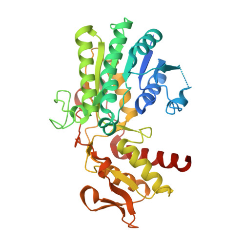 | |
UniProt & NIH Common Fund Data Resources | |||||
Find proteins for O60547 (Homo sapiens) Explore O60547 Go to UniProtKB: O60547 | |||||
PHAROS: O60547 GTEx: ENSG00000112699 | |||||
Entity Groups | |||||
| Sequence Clusters | 30% Identity50% Identity70% Identity90% Identity95% Identity100% Identity | ||||
| UniProt Group | O60547 | ||||
Sequence AnnotationsExpand | |||||
| |||||
| Ligands 2 Unique | |||||
|---|---|---|---|---|---|
| ID | Chains | Name / Formula / InChI Key | 2D Diagram | 3D Interactions | |
| NDP Query on NDP | E [auth A], G [auth B], I [auth C], K [auth D] | NADPH DIHYDRO-NICOTINAMIDE-ADENINE-DINUCLEOTIDE PHOSPHATE C21 H30 N7 O17 P3 ACFIXJIJDZMPPO-NNYOXOHSSA-N |  | ||
| GDP Query on GDP | F [auth A], H [auth B], J [auth C], L [auth D] | GUANOSINE-5'-DIPHOSPHATE C10 H15 N5 O11 P2 QGWNDRXFNXRZMB-UUOKFMHZSA-N |  | ||
| Length ( Å ) | Angle ( ˚ ) |
|---|---|
| a = 88.595 | α = 90 |
| b = 122.064 | β = 90 |
| c = 139.403 | γ = 90 |
| Software Name | Purpose |
|---|---|
| CNS | refinement |
| HKL-2000 | data reduction |
| SCALEPACK | data scaling |
| CNS | phasing |
RCSB PDB (citation) is hosted by
RCSB PDB is a member of the

