1SQN

Progesterone Receptor Ligand Binding Domain with bound Norethindrone


Experimental Data Snapshot

  • Method: X-RAY DIFFRACTION
  • Resolution: 1.45 Å
  • R-Value Free: 0.216 
  • R-Value Work: 0.186 
  • R-Value Observed: 0.188 

wwPDB Validation   3D Report Full Report


Ligand Structure Quality Assessment 


This is version 1.4 of the entry. See complete history


Literature

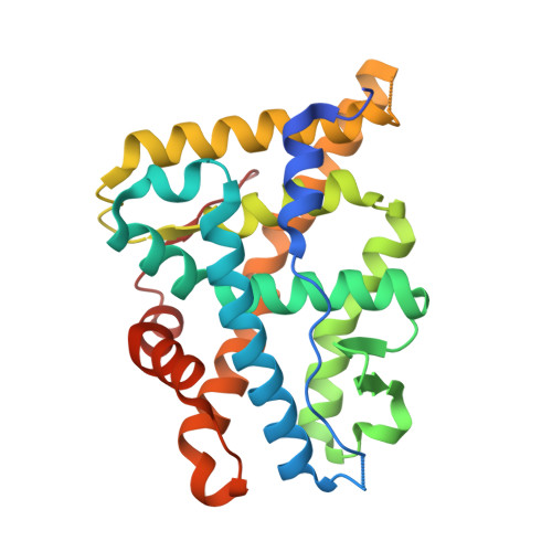
Progesterone receptor ligand binding pocket flexibility: crystal structures of the norethindrone and mometasone furoate complexes

Madauss, K.P.Deng, J.-S.Austin, R.J.H.Lambert, M.H.McLay, I.Pritchard, J.Short, S.A.Stewart, E.L.Uings, I.J.Williams, S.P.

(2004) J Med Chem 47: 3381-3387

  • DOI: https://doi.org/10.1021/jm030640n
  • Primary Citation of Related Structures:  
    1SQN, 1SR7

  • PubMed Abstract: 

    Although progesterone, the natural ligand of the progesterone receptor (PR), has a hydrogen atom at the 17alpha position, other potent steroid agonists such as norethindrone and mometasone furoate have larger substituents at this position that are accommodated by the PR ligand binding pocket. Crystallographic analysis of PR ligand binding domain complexes clearly demonstrated that these moieties were accommodated by local shifts of the protein main chain and by adoption of alternative side chain rotamer conformations of ligand-proximal amino acids. These conformational changes imparted a ligand-specific volume to the binding pocket, from 490 A3 in the metribolone complex to 520 A3 in the norethindrone complex, 565 A3 in the progesterone complex, and 730 A3 in the mometasone furoate complex. Despite these marked alterations in binding pocket volume, critical interactions essential for establishment of an active AF2 conformation were maintained.


  • Organizational Affiliation

    GlaxoSmithKline Inc., 5 Moore Drive, Research Triangle Park, North Carolina 27709, USA.


Macromolecules
Find similar proteins by:  (by identity cutoff)  |  3D Structure
Entity ID: 1
MoleculeChains Sequence LengthOrganismDetailsImage
progesterone receptor
A, B
261Homo sapiensMutation(s): 0 
Gene Names: HUMPGRR
UniProt & NIH Common Fund Data Resources
Find proteins for P06401 (Homo sapiens)
Explore P06401 
Go to UniProtKB:  P06401
PHAROS:  P06401
GTEx:  ENSG00000082175 
Entity Groups  
Sequence Clusters30% Identity50% Identity70% Identity90% Identity95% Identity100% Identity
UniProt GroupP06401
Sequence Annotations
Expand
  • Reference Sequence
Small Molecules
Ligands 1 Unique
IDChains Name / Formula / InChI Key2D Diagram3D Interactions
NDR
Query on NDR

Download Ideal Coordinates CCD File 
C [auth A],
D [auth B]
(14beta,17alpha)-17-ethynyl-17-hydroxyestr-4-en-3-one
C20 H26 O2
VIKNJXKGJWUCNN-XGXHKTLJSA-N
Binding Affinity Annotations 
IDSourceBinding Affinity
NDR BindingDB:  1SQN Ki: 1.9 (nM) from 1 assay(s)
Kd: 0.4 (nM) from 1 assay(s)
IC50: 3.2 (nM) from 1 assay(s)
EC50: 2.2 (nM) from 1 assay(s)
Binding MOAD:  1SQN Kd: 0.4 (nM) from 1 assay(s)
PDBBind:  1SQN Kd: 0.4 (nM) from 1 assay(s)
Experimental Data & Validation

Experimental Data

  • Method: X-RAY DIFFRACTION
  • Resolution: 1.45 Å
  • R-Value Free: 0.216 
  • R-Value Work: 0.186 
  • R-Value Observed: 0.188 
  • Space Group: P 1 21 1
Unit Cell:
Length ( Å )Angle ( ˚ )
a = 57.753α = 90
b = 64.142β = 95.84
c = 70.138γ = 90
Software Package:
Software NamePurpose
ADSCdata collection
SCALEPACKdata scaling
AMoREphasing
REFMACrefinement

Structure Validation

View Full Validation Report


Ligand Structure Quality Assessment 


Entry History 

Deposition Data

Revision History  (Full details and data files)

  • Version 1.0: 2004-07-27
    Type: Initial release
  • Version 1.1: 2008-04-29
    Changes: Version format compliance
  • Version 1.2: 2011-07-13
    Changes: Version format compliance
  • Version 1.3: 2018-01-31
    Changes: Experimental preparation
  • Version 1.4: 2023-08-23
    Changes: Data collection, Database references, Derived calculations, Refinement description