1RFG

Crystal Structure of Human Purine Nucleoside Phosphorylase Complexed with Guanosine


Experimental Data Snapshot

  • Method: X-RAY DIFFRACTION
  • Resolution: 2.90 Å
  • R-Value Free: 0.267 
  • R-Value Work: 0.206 
  • R-Value Observed: 0.206 

wwPDB Validation   3D Report Full Report


Ligand Structure Quality Assessment 


This is version 1.3 of the entry. See complete history


Literature

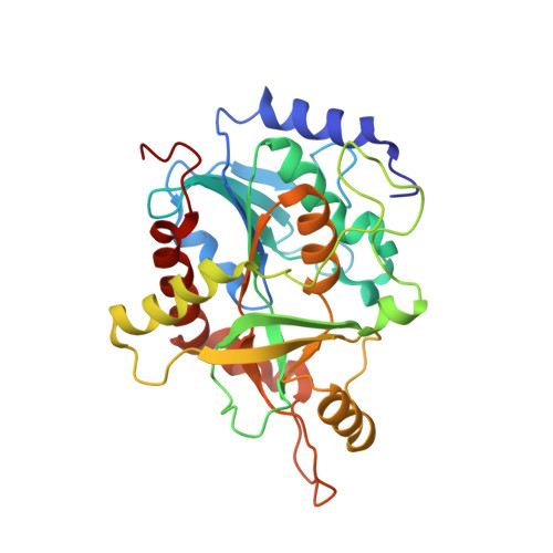
Structure of human PNP complexed with ligands.

Canduri, F.Silva, R.G.dos Santos, D.M.Palma, M.S.Basso, L.A.Santos, D.S.de Azevedo, W.F.

(2005) Acta Crystallogr D Biol Crystallogr 61: 856-862

  • DOI: https://doi.org/10.1107/S0907444905005421
  • Primary Citation of Related Structures:  
    1RFG, 1V41, 1V45

  • PubMed Abstract: 

    Purine nucleoside phosphorylase (PNP) is a key enzyme in the purine-salvage pathway, which allows cells to utilize preformed bases and nucleosides in order to synthesize nucleotides. PNP is specific for purine nucleosides in the beta-configuration and exhibits a strong preference for purines containing a 6-keto group and ribosyl-containing nucleosides relative to the corresponding analogues. PNP was crystallized in complex with ligands and data collection was performed using synchrotron radiation. This work reports the structure of human PNP in complex with guanosine (at 2.80 A resolution), 3'-deoxyguanosine (at 2.86 A resolution) and 8-azaguanine (at 2.85 A resolution). These structures were compared with the PNP-guanine, PNP-inosine and PNP-immucillin-H complexes solved previously.


  • Organizational Affiliation

    Programa de Pós-Graduação em Biofísica Molecular, Departamento de Física, UNESP, São José do Rio Preto, SP 15054-000, Brazil.


Macromolecules
Find similar proteins by:  (by identity cutoff)  |  3D Structure
Entity ID: 1
MoleculeChains Sequence LengthOrganismDetailsImage
Purine nucleoside phosphorylaseA [auth E]288Homo sapiensMutation(s): 0 
Gene Names: NPPNP
EC: 2.4.2.1
UniProt & NIH Common Fund Data Resources
Find proteins for P00491 (Homo sapiens)
Explore P00491 
Go to UniProtKB:  P00491
PHAROS:  P00491
GTEx:  ENSG00000198805 
Entity Groups  
Sequence Clusters30% Identity50% Identity70% Identity90% Identity95% Identity100% Identity
UniProt GroupP00491
Sequence Annotations
Expand
  • Reference Sequence
Experimental Data & Validation

Experimental Data

  • Method: X-RAY DIFFRACTION
  • Resolution: 2.90 Å
  • R-Value Free: 0.267 
  • R-Value Work: 0.206 
  • R-Value Observed: 0.206 
  • Space Group: H 3 2
Unit Cell:
Length ( Å )Angle ( ˚ )
a = 139.38α = 90
b = 139.38β = 90
c = 160.4γ = 120
Software Package:
Software NamePurpose
MOSFLMdata reduction
SCALAdata scaling
AMoREphasing
X-PLORrefinement
CCP4data scaling

Structure Validation

View Full Validation Report


Ligand Structure Quality Assessment 


Entry History 

Deposition Data

Revision History  (Full details and data files)

  • Version 1.0: 2004-12-14
    Type: Initial release
  • Version 1.1: 2008-04-29
    Changes: Version format compliance
  • Version 1.2: 2011-07-13
    Changes: Derived calculations, Version format compliance
  • Version 1.3: 2023-08-23
    Changes: Data collection, Database references, Derived calculations, Refinement description