1QUN

X-RAY STRUCTURE OF THE FIMC-FIMH CHAPERONE ADHESIN COMPLEX FROM UROPATHOGENIC E.COLI


Experimental Data Snapshot

  • Method: X-RAY DIFFRACTION
  • Resolution: 2.80 Å
  • R-Value Free: 0.268 
  • R-Value Work: 0.240 

wwPDB Validation   3D Report Full Report


This is version 1.2 of the entry. See complete history


Literature

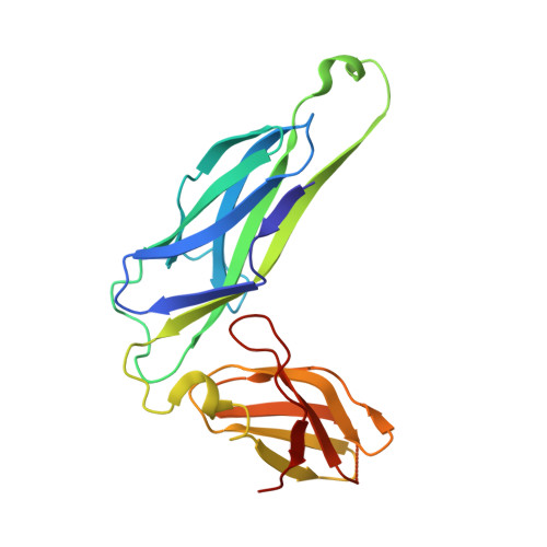
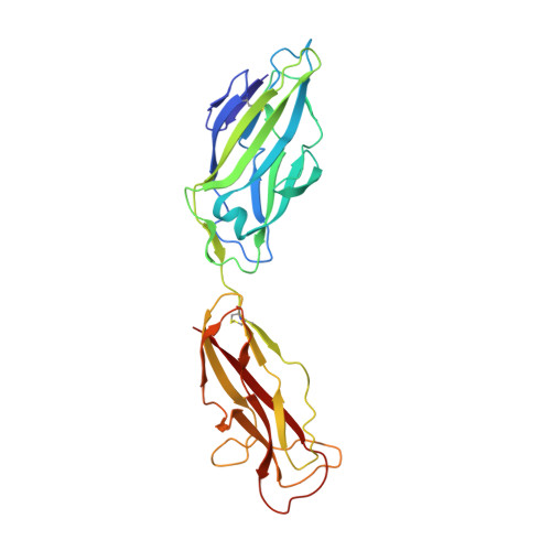
X-ray structure of the FimC-FimH chaperone-adhesin complex from uropathogenic Escherichia coli.

Choudhury, D.Thompson, A.Stojanoff, V.Langermann, S.Pinkner, J.Hultgren, S.J.Knight, S.D.

(1999) Science 285: 1061-1066

  • DOI: https://doi.org/10.1126/science.285.5430.1061
  • Primary Citation of Related Structures:  
    1QUN

  • PubMed Abstract: 

    Type 1 pili-adhesive fibers expressed in most members of the Enterobacteriaceae family-mediate binding to mannose receptors on host cells through the FimH adhesin. Pilus biogenesis proceeds by way of the chaperone/usher pathway. The x-ray structure of the FimC-FimH chaperone-adhesin complex from uropathogenic Escherichia coli at 2.5 angstrom resolution reveals the basis for carbohydrate recognition and for pilus assembly. The carboxyl-terminal pilin domain of FimH has an immunoglobulin-like fold, except that the seventh strand is missing, leaving part of the hydrophobic core exposed. A donor strand complementation mechanism in which the chaperone donates a strand to complete the pilin domain explains the basis for both chaperone function and pilus biogenesis.


  • Organizational Affiliation

    Department of Molecular Biology, Uppsala Biomedical Center, Swedish University of Agricultural Sciences, Box 590, S-753 24 Uppsala, Sweden.


Macromolecules
Find similar proteins by:  (by identity cutoff)  |  3D Structure
Entity ID: 1
MoleculeChains Sequence LengthOrganismDetailsImage
PAPD-LIKE CHAPERONE FIMC
A, C, E, G, I
A, C, E, G, I, K, M, O
205Escherichia coliMutation(s): 0 
UniProt
Find proteins for P31697 (Escherichia coli (strain K12))
Explore P31697 
Go to UniProtKB:  P31697
Entity Groups  
Sequence Clusters30% Identity50% Identity70% Identity90% Identity95% Identity100% Identity
UniProt GroupP31697
Sequence Annotations
Expand
  • Reference Sequence
Find similar proteins by:  (by identity cutoff)  |  3D Structure
Entity ID: 2
MoleculeChains Sequence LengthOrganismDetailsImage
MANNOSE-SPECIFIC ADHESIN FIMH
B, D, F, H, J
B, D, F, H, J, L, N, P
279Escherichia coliMutation(s): 0 
UniProt
Find proteins for P08191 (Escherichia coli (strain K12))
Explore P08191 
Go to UniProtKB:  P08191
Entity Groups  
Sequence Clusters30% Identity50% Identity70% Identity90% Identity95% Identity100% Identity
UniProt GroupP08191
Sequence Annotations
Expand
  • Reference Sequence
Experimental Data & Validation

Experimental Data

  • Method: X-RAY DIFFRACTION
  • Resolution: 2.80 Å
  • R-Value Free: 0.268 
  • R-Value Work: 0.240 
  • Space Group: C 1 2 1
Unit Cell:
Length ( Å )Angle ( ˚ )
a = 139.41α = 90
b = 139.57β = 90
c = 215.72γ = 90
Software Package:
Software NamePurpose
MOSFLMdata reduction
SCALAdata scaling
SOLVEphasing
REFMACrefinement
CCP4data scaling

Structure Validation

View Full Validation Report


Entry History 

Deposition Data

Revision History  (Full details and data files)

  • Version 1.0: 1999-08-31
    Type: Initial release
  • Version 1.1: 2008-04-27
    Changes: Version format compliance
  • Version 1.2: 2011-07-13
    Changes: Version format compliance