Three dimensional structure of Mammalian tachykinin Peptide neurokinin B bound to lipid micelles.
Mantha, A.K., Chandrashekar, I.R., Baquer, N.Z., Cowsik, S.M.(2004) J Biomol Struct Dyn 22: 137-148
- PubMed: 15317475
- DOI: https://doi.org/10.1080/07391102.2004.10506990
- Primary Citation of Related Structures:
1P9F - PubMed Abstract:
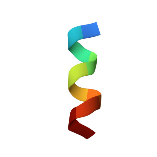
Neurokinin B (NKB), a decapeptide of mammalian origin exhibits a variety of biological activities such as regulatory functions in reproduction, pre-eclampsia and neuroprotection in Alzheimer's disease. In order to gain insight into structure-function relationship, three-dimensional structure of NKB has been investigated using CD spectropolarimetry and two-dimensional proton nuclear magnetic resonance (2D 1H-NMR) spectroscopy in aqueous and membrane mimetic solvents. Unambiguous NMR assignments of resonances have been made with the aid of correlation spectroscopy (DQF-COSY and TOCSY) experiments and Nuclear Overhauser Effect Spectroscopy (NOESY) experiments. Distance constraints obtained from the NMR data have been used to generate a family of structures, which have been refined using restrained energy minimization and dynamics. Our data show that a helical structure is induced in NKB, in presence of perdeuterated dodecyl phosphocholine (DPC) micelles, a membrane model system. Further, the conformation adopted by NKB in presence of DPC micelles represents a structural motif typical of neurokinin-3 selective agonists.
Organizational Affiliation:
School of Life Sciences, Jawaharlal Nehru University, New Delhi--110 067, India.