1P7H

Structure of NFAT1 bound as a dimer to the HIV-1 LTR kB element


Experimental Data Snapshot

  • Method: X-RAY DIFFRACTION
  • Resolution: 2.60 Å
  • R-Value Free: 0.265 
  • R-Value Work: 0.231 
  • R-Value Observed: 0.231 

wwPDB Validation   3D Report Full Report


This is version 1.4 of the entry. See complete history


Literature

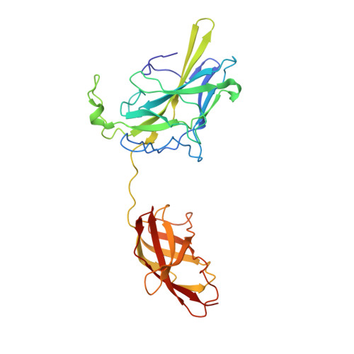
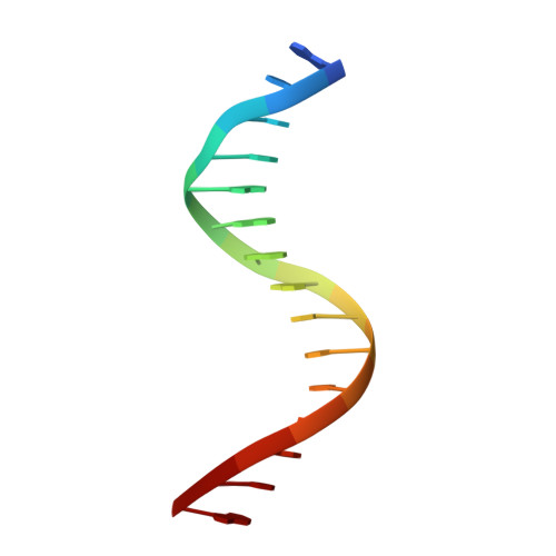
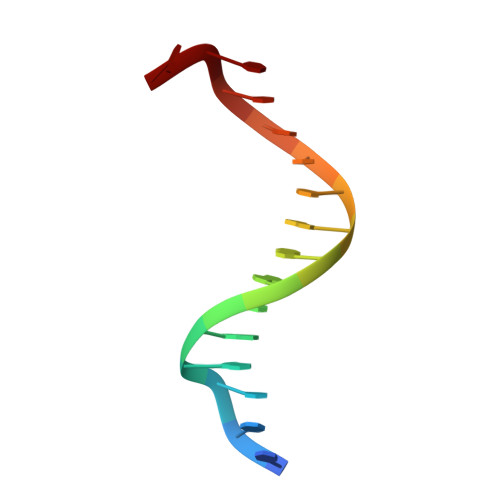
Structure of NFAT1 bound as a dimer to the HIV-1 LTR kappa B element

Giffin, M.J.Stroud, J.C.Bates, D.L.von Koenig, K.D.Hardin, J.Chen, L.

(2003) Nat Struct Biol 10: 800-806

  • DOI: https://doi.org/10.1038/nsb981
  • Primary Citation of Related Structures:  
    1P7H

  • PubMed Abstract: 

    DNA binding by NFAT1 as a dimer has been implicated in the activation of host and viral genes. Here we report a crystal structure of NFAT1 bound cooperatively as a dimer to the highly conserved kappa B site from the human immunodeficiency virus 1 (HIV-1) long terminal repeat (LTR). This structure reveals a new mode of dimerization and protein-DNA recognition by the Rel homology region (RHR) of NFAT1. The two NFAT1 monomers form a complete circle around the kappa B DNA through protein-protein interactions mediated by both their N- and C-terminal subdomains. The major dimer interface, formed by the C-terminal domain, is asymmetric and substantially different from the symmetric dimer interface seen in other Rel family proteins. Comparison to other NFAT structures, including NFAT5 and the NFAT1-Fos-Jun-ARRE2 complex, reveals that NFAT1 adopts different conformations and its protein surfaces mediate distinct protein-protein interactions in the context of different DNA sites.


  • Organizational Affiliation

    Department of Chemistry and Biochemistry, University of Colorado at Boulder, Boulder, Colorado 80309-0215, USA.


Macromolecules

Find similar proteins by:  (by identity cutoff)  |  3D Structure
Entity ID: 3
MoleculeChains Sequence LengthOrganismDetailsImage
Nuclear factor of activated T-cells, cytoplasmic 2E [auth L],
F [auth M],
G [auth N],
H [auth O]
286Homo sapiensMutation(s): 0 
Gene Names: NFATC2 OR NFAT1 OR NFATP
UniProt & NIH Common Fund Data Resources
Find proteins for Q13469 (Homo sapiens)
Explore Q13469 
Go to UniProtKB:  Q13469
PHAROS:  Q13469
GTEx:  ENSG00000101096 
Entity Groups  
Sequence Clusters30% Identity50% Identity70% Identity90% Identity95% Identity100% Identity
UniProt GroupQ13469
Sequence Annotations
Expand
  • Reference Sequence

Find similar nucleic acids by:  Sequence   |   3D Structure  

Entity ID: 1
MoleculeChains LengthOrganismImage
5'-D(*AP*AP*TP*GP*GP*GP*GP*AP*CP*TP*TP*TP*CP*CP*A)-3'
A, C
15N/A
Sequence Annotations
Expand
  • Reference Sequence

Find similar nucleic acids by:  Sequence   |   3D Structure  

Entity ID: 2
MoleculeChains LengthOrganismImage
5'-D(*TP*TP*TP*GP*GP*AP*AP*AP*GP*TP*CP*CP*CP*CP*A)-3'
B, D
15N/A
Sequence Annotations
Expand
  • Reference Sequence
Experimental Data & Validation

Experimental Data

  • Method: X-RAY DIFFRACTION
  • Resolution: 2.60 Å
  • R-Value Free: 0.265 
  • R-Value Work: 0.231 
  • R-Value Observed: 0.231 
  • Space Group: P 1
Unit Cell:
Length ( Å )Angle ( ˚ )
a = 74.107α = 71.2
b = 80.321β = 78.97
c = 80.308γ = 78.94
Software Package:
Software NamePurpose
CNSrefinement
Adxvdata processing
SCALEPACKdata scaling
CNSphasing

Structure Validation

View Full Validation Report


Entry History 

Deposition Data

Revision History  (Full details and data files)

  • Version 1.0: 2003-09-23
    Type: Initial release
  • Version 1.1: 2008-04-29
    Changes: Version format compliance
  • Version 1.2: 2011-07-13
    Changes: Version format compliance
  • Version 1.3: 2017-10-11
    Changes: Refinement description
  • Version 1.4: 2024-02-14
    Changes: Data collection, Database references