1OAK

CRYSTAL STRUCTURE OF THE VON WILLEBRAND FACTOR (VWF) A1 DOMAIN IN COMPLEX WITH THE FUNCTION BLOCKING NMC-4 FAB


Experimental Data Snapshot

  • Method: X-RAY DIFFRACTION
  • Resolution: 2.20 Å
  • R-Value Free: 0.274 
  • R-Value Work: 0.201 
  • R-Value Observed: 0.201 

wwPDB Validation   3D Report Full Report


This is version 1.2 of the entry. See complete history


Literature

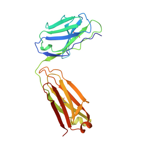
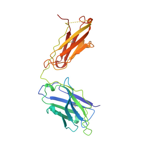
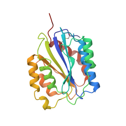
Crystal structure of the von Willebrand factor A1 domain in complex with the function blocking NMC-4 Fab.

Celikel, R.Varughese, K.I.MadhusudanYoshioka, A.Ware, J.Ruggeri, Z.M.

(1998) Nat Struct Biol 5: 189-194

  • DOI: https://doi.org/10.1038/nsb0398-189
  • Primary Citation of Related Structures:  
    1OAK

  • PubMed Abstract: 

    The presence of one or more copies of von Willebrand factor type A domains identifies a superfamily of proteins usually involved in biological processes controlled by specific molecular interactions, often adhesive in nature. We have solved the crystal structure of the prototypic von Willebrand factor A1 domain, essential for the antihemorrhagic activity of platelets, in complex with the function blocking antibody, NMC-4, at 2.2 A resolution. This has led to the recognition of a putative binding groove for the platelet receptor, glycoprotein Ib alpha, formed by two adjacent alpha-helices and a beta-strand. The structure also shows a contact interface between A1 domain pairs, suggesting a hypothetical mechanism for the regulation of protein assembly and heterologous ligand binding mediated by homophilic interactions of type A domains.


  • Organizational Affiliation

    Roon Research Center for Arteriosclerosis and Thrombosis, The Scripps Research Institute, La Jolla, California 92037, USA.


Macromolecules
Find similar proteins by:  (by identity cutoff)  |  3D Structure
Entity ID: 1
MoleculeChains Sequence LengthOrganismDetailsImage
NMC-4 IGG1A [auth L]212Mus musculusMutation(s): 0 
Entity Groups  
Sequence Clusters30% Identity50% Identity70% Identity90% Identity95% Identity100% Identity
Sequence Annotations
Expand
  • Reference Sequence
Find similar proteins by:  (by identity cutoff)  |  3D Structure
Entity ID: 2
MoleculeChains Sequence LengthOrganismDetailsImage
NMC-4 IGG1B [auth H]223Mus musculusMutation(s): 0 
UniProt
Find proteins for P01868 (Mus musculus)
Explore P01868 
Go to UniProtKB:  P01868
Entity Groups  
Sequence Clusters30% Identity50% Identity70% Identity90% Identity95% Identity100% Identity
UniProt GroupP01868
Sequence Annotations
Expand
  • Reference Sequence
Find similar proteins by:  (by identity cutoff)  |  3D Structure
Entity ID: 3
MoleculeChains Sequence LengthOrganismDetailsImage
VON WILLEBRAND FACTORC [auth A]196Homo sapiensMutation(s): 0 
UniProt & NIH Common Fund Data Resources
Find proteins for P04275 (Homo sapiens)
Explore P04275 
Go to UniProtKB:  P04275
PHAROS:  P04275
GTEx:  ENSG00000110799 
Entity Groups  
Sequence Clusters30% Identity50% Identity70% Identity90% Identity95% Identity100% Identity
UniProt GroupP04275
Sequence Annotations
Expand
  • Reference Sequence
Experimental Data & Validation

Experimental Data

  • Method: X-RAY DIFFRACTION
  • Resolution: 2.20 Å
  • R-Value Free: 0.274 
  • R-Value Work: 0.201 
  • R-Value Observed: 0.201 
  • Space Group: C 1 2 1
Unit Cell:
Length ( Å )Angle ( ˚ )
a = 208.2α = 90
b = 61.9β = 108.6
c = 72.9γ = 90
Software Package:
Software NamePurpose
DENZOdata reduction
SCALEPACKdata scaling
PHASESphasing
X-PLORmodel building
X-PLORrefinement
X-PLORphasing

Structure Validation

View Full Validation Report


Entry History 

Deposition Data

Revision History  (Full details and data files)

  • Version 1.0: 1998-10-21
    Type: Initial release
  • Version 1.1: 2008-03-24
    Changes: Version format compliance
  • Version 1.2: 2011-07-13
    Changes: Version format compliance