1M4K

Crystal structure of the human natural killer cell activator receptor KIR2DS2 (CD158j)


Experimental Data Snapshot

  • Method: X-RAY DIFFRACTION
  • Resolution: 2.30 Å
  • R-Value Free: 0.247 
  • R-Value Work: 0.221 
  • R-Value Observed: 0.230 

wwPDB Validation   3D Report Full Report


This is version 1.3 of the entry. See complete history


Literature

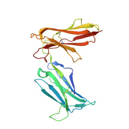
Crystal structure of the human natural killer cell activating receptor KIR2DS2 (CD158j)

Saulquin, X.Gastinel, L.N.Vivier, E.

(2003) J Exp Med 197: 933-938

  • DOI: https://doi.org/10.1084/jem.20021624
  • Primary Citation of Related Structures:  
    1M4K

  • PubMed Abstract: 

    Killer cell Ig-like receptors (KIRs) regulate the function of human natural killer and T cell subsets. A feature of the KIR locus is the clustering of homologous genes encoding for inhibitory and activating KIR. Inhibitory and activating KIR differ for ligand specificities and/or affinities. In particular, we show here with KIR tetramers that activating KIR2DS2 does not bind HLA-Cw3 molecules recognized by inhibitory KIR2DL2, despite 99% extracellular amino acid identity. We also report the 2.3-A structure of KIR2DS2, which reveals subtle displacements of two residues (Tyr45 and Gln71) involved in the interaction of KIR2DL2 with HLA-Cw3. These results show that KIR molecules cannot tolerate any variability in their three-dimensional structure without altering their MHC class I recognition capacities. Therefore, the mode of recognition used by KIR largely differs from the conformational changes that characterize T cell receptor or NKG2D interaction with their respective ligands.


  • Organizational Affiliation

    Centre d'Immunologie INSERM-CNRS, de Marseille Luminy, Case 906, Parc Scientifique de Luminy, 13288 Marseille Cedex 09, France.


Macromolecules
Find similar proteins by:  (by identity cutoff)  |  3D Structure
Entity ID: 1
MoleculeChains Sequence LengthOrganismDetailsImage
KILLER CELL IMMUNOGLOBULIN-LIKE RECEPTOR 2DS2200Homo sapiensMutation(s): 0 
UniProt
Find proteins for P43631 (Homo sapiens)
Explore P43631 
Go to UniProtKB:  P43631
Entity Groups  
Sequence Clusters30% Identity50% Identity70% Identity90% Identity95% Identity100% Identity
UniProt GroupP43631
Sequence Annotations
Expand
  • Reference Sequence
Small Molecules
Experimental Data & Validation

Experimental Data

  • Method: X-RAY DIFFRACTION
  • Resolution: 2.30 Å
  • R-Value Free: 0.247 
  • R-Value Work: 0.221 
  • R-Value Observed: 0.230 
  • Space Group: P 31 2 1
Unit Cell:
Length ( Å )Angle ( ˚ )
a = 97.535α = 90
b = 97.535β = 90
c = 54.375γ = 120
Software Package:
Software NamePurpose
DENZOdata reduction
SCALEPACKdata scaling
AMoREphasing
CNSrefinement

Structure Validation

View Full Validation Report


Entry History 

Deposition Data

Revision History  (Full details and data files)

  • Version 1.0: 2003-04-15
    Type: Initial release
  • Version 1.1: 2008-04-28
    Changes: Version format compliance
  • Version 1.2: 2011-07-13
    Changes: Version format compliance
  • Version 1.3: 2024-04-03
    Changes: Data collection, Database references, Derived calculations, Refinement description