1LQ8

Crystal structure of cleaved protein C inhibitor


Experimental Data Snapshot

  • Method: X-RAY DIFFRACTION
  • Resolution: 2.40 Å
  • R-Value Free: 0.279 
  • R-Value Work: 0.222 

wwPDB Validation   3D Report Full Report


Ligand Structure Quality Assessment 


This is version 2.1 of the entry. See complete history


Literature

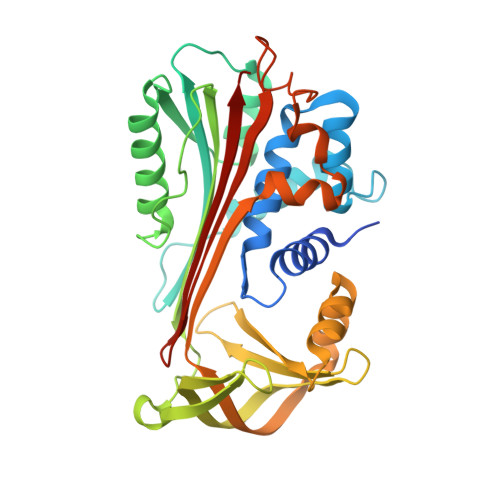
Crystal Structure of Protein C Inhibitor Provides Insights into Hormone Binding and Heparin Activation

Huntington, J.A.Kjellberg, M.Stenflo, J.

(2003) Structure 11: 205-211

  • DOI: https://doi.org/10.1016/s0969-2126(02)00944-9
  • Primary Citation of Related Structures:  
    1LQ8

  • PubMed Abstract: 

    Protein C inhibitor (PCI) is a member of the serpin family that has many biological functions. In blood it acts as a procoagulant, and, in the seminal vesicles, it is required for spermatogenesis. The activity of PCI is affected by heparin binding in a manner unique among the heparin binding serpins, and, in addition, PCI binds hydrophobic hormones with apparent specificity for retinoids. Here we present the 2.4 A crystallographic structure of reactive center loop (RCL) cleaved PCI. A striking feature of the structure is a two-turn N-terminal shortening of helix A, which creates a large hydrophobic pocket that docking studies indicate to be the retinoid binding site. On the basis of surface electrostatic properties, a novel mechanism for heparin activation is proposed.


  • Organizational Affiliation

    Department of Haematology, Division of Structural Medicine, University of Cambridge, Cambridge Institute for Medical Research, Wellcome Trust/MRC Building, Hills Road, CB2 2XY, Cambridge, United Kingdom. jah52@cam.ac.uk


Macromolecules
Find similar proteins by:  (by identity cutoff)  |  3D Structure
Entity ID: 1
MoleculeChains Sequence LengthOrganismDetailsImage
plasma serine protease inhibitor
A, C, E, G
346Homo sapiensMutation(s): 0 
UniProt & NIH Common Fund Data Resources
Find proteins for P05154 (Homo sapiens)
Explore P05154 
Go to UniProtKB:  P05154
PHAROS:  P05154
GTEx:  ENSG00000188488 
Entity Groups  
Sequence Clusters30% Identity50% Identity70% Identity90% Identity95% Identity100% Identity
UniProt GroupP05154
Sequence Annotations
Expand
  • Reference Sequence
Find similar proteins by:  (by identity cutoff)  |  3D Structure
Entity ID: 2
MoleculeChains Sequence LengthOrganismDetailsImage
plasma serine protease inhibitor
B, D, F, H
31Homo sapiensMutation(s): 0 
UniProt & NIH Common Fund Data Resources
Find proteins for P05154 (Homo sapiens)
Explore P05154 
Go to UniProtKB:  P05154
PHAROS:  P05154
GTEx:  ENSG00000188488 
Entity Groups  
Sequence Clusters30% Identity50% Identity70% Identity90% Identity95% Identity100% Identity
UniProt GroupP05154
Sequence Annotations
Expand
  • Reference Sequence
Oligosaccharides

Help

Entity ID: 3
MoleculeChains Length2D Diagram Glycosylation3D Interactions
2-acetamido-2-deoxy-alpha-D-glucopyranose-(1-4)-2-acetamido-2-deoxy-beta-D-glucopyranose
I, M
2N-Glycosylation
Glycosylation Resources
GlyTouCan:  G07375KG
GlyCosmos:  G07375KG
GlyGen:  G07375KG
Entity ID: 4
MoleculeChains Length2D Diagram Glycosylation3D Interactions
2-acetamido-2-deoxy-beta-D-glucopyranose-(1-4)-2-acetamido-2-deoxy-beta-D-glucopyranose
J, K
2N-Glycosylation
Glycosylation Resources
GlyTouCan:  G42666HT
GlyCosmos:  G42666HT
GlyGen:  G42666HT
Entity ID: 5
MoleculeChains Length2D Diagram Glycosylation3D Interactions
alpha-D-mannopyranose-(1-3)-[alpha-D-mannopyranose-(1-6)]alpha-D-mannopyranose
L
3N/A
Glycosylation Resources
GlyTouCan:  G00619DD
GlyCosmos:  G00619DD
GlyGen:  G00619DD
Experimental Data & Validation

Experimental Data

  • Method: X-RAY DIFFRACTION
  • Resolution: 2.40 Å
  • R-Value Free: 0.279 
  • R-Value Work: 0.222 
  • Space Group: P 1 21 1
Unit Cell:
Length ( Å )Angle ( ˚ )
a = 55.2α = 90
b = 244.03β = 91.98
c = 66.4γ = 90
Software Package:
Software NamePurpose
HKL-2000data collection
SCALEPACKdata scaling
MOLREPphasing
CNSrefinement
HKL-2000data reduction

Structure Validation

View Full Validation Report


Ligand Structure Quality Assessment 


Entry History 

Deposition Data

Revision History  (Full details and data files)

  • Version 1.0: 2003-02-11
    Type: Initial release
  • Version 1.1: 2007-10-16
    Changes: Version format compliance
  • Version 1.2: 2011-07-13
    Changes: Derived calculations, Version format compliance
  • Version 2.0: 2020-07-29
    Type: Remediation
    Reason: Carbohydrate remediation
    Changes: Advisory, Atomic model, Data collection, Derived calculations, Structure summary
  • Version 2.1: 2023-08-16
    Changes: Advisory, Data collection, Database references, Refinement description, Structure summary