Transforming growth factor beta 1: three-dimensional structure in solution and comparison with the X-ray structure of transforming growth factor beta 2.
Hinck, A.P., Archer, S.J., Qian, S.W., Roberts, A.B., Sporn, M.B., Weatherbee, J.A., Tsang, M.L., Lucas, R., Zhang, B.L., Wenker, J., Torchia, D.A.(1996) Biochemistry 35: 8517-8534
- PubMed: 8679613
- DOI: https://doi.org/10.1021/bi9604946
- Primary Citation of Related Structures:
1KLA, 1KLC, 1KLD - PubMed Abstract:
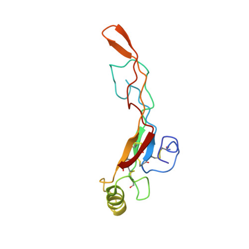
The three-dimensional solution structure of human transforming growth factor beta 1 (TGF-beta 1) has been determined using multinuclear magnetic resonance spectroscopy and a hybrid distance geometry/ simulated annealing algorithm. It represents one of the first examples of a mammalian protein structure that has been solved by isotopic labeling of the protein in a eukaryotic cell line and multinuclear NMR spectroscopy. The solution structure of the 25 kDa disulfide-linked TGF-beta 1 homodimer was calculated from over 3200 distance and dihedral angle restraints. The final ensemble of 33 accepted structures had no NOE or dihedral angle violations greater than 0.30 A and 5.0 degrees, respectively. The RMSD of backbone atoms for the ensemble of 33 structures relative to their mean structure was 1.1 A when all residues were used in the alignment and 0.7 A when loop regions were omitted. The solution structure of TGF-beta 1 follows two independently determined crystal structures of TGF-beta 2 (Daopin et al., 1992, 1993; Schlunegger & Grütter, 1992, 1993), providing the first opportunity to examine structural differences between the two isoforms at the molecular level. Although the structures are very similar, with an RMSD in backbone atom positions of 1.4 A when loop regions are omitted in the alignment and 1.9 A when all residues are considered, there are several notable differences in structure and flexibility which may be related to function. The clearest example of these is in the beta-turn from residues 69-72: the turn type found in the solution structure of TGF-beta 1 falls into the category of type II, whereas that present in the X-ray crystal structure of TGF-beta 2 is more consistent with a type I turn conformation. This may be of functional significance as studies using TGF-beta chimeras and deletion mutants indicate that this portion of the molecule may be important in receptor binding.
Organizational Affiliation:
Molecular Structural Biology Unit, National Institute of Dental Research, National Institutes of Health, Bethesda, Maryland 20892-4326, USA.