1IPC

CRYSTAL STRUCTURE OF EUKARYOTIC INITIATION FACTOR 4E COMPLEXED WITH 7-METHYL GTP


Experimental Data Snapshot

  • Method: X-RAY DIFFRACTION
  • Resolution: 2.00 Å
  • R-Value Free: 0.228 
  • R-Value Work: 0.192 

wwPDB Validation   3D Report Full Report


Ligand Structure Quality Assessment 


This is version 1.4 of the entry. See complete history


Literature

Crystal structures of 7-methylguanosine 5'-triphosphate (m(7)GTP)- and P(1)-7-methylguanosine-P(3)-adenosine-5',5'-triphosphate (m(7)GpppA)-bound human full-length eukaryotic initiation factor 4E: biological importance of the C-terminal flexible region

Tomoo, K.Shen, X.Okabe, K.Nozoe, Y.Fukuhara, S.Morino, S.Ishida, T.Taniguchi, T.Hasegawa, H.Terashima, A.Sasaki, M.Katsuya, Y.Kitamura, K.Miyoshi, H.Ishikawa, M.Miura, K.

(2002) Biochem J 362: 539-544

  • DOI: https://doi.org/10.1042/0264-6021:3620539
  • Primary Citation of Related Structures:  
    1IPB, 1IPC

  • PubMed Abstract: 

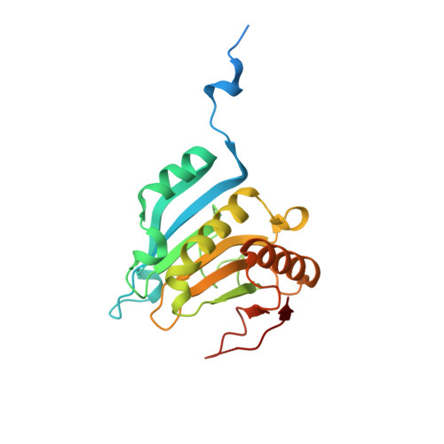
    The crystal structures of the full-length human eukaryotic initiation factor (eIF) 4E complexed with two mRNA cap analogues [7-methylguanosine 5'-triphosphate (m(7)GTP) and P(1)-7-methylguanosine-P(3)-adenosine-5',5'-triphosphate (m(7)GpppA)] were determined at 2.0 A resolution (where 1 A=0.1 nm). The flexibility of the C-terminal loop region of eIF4E complexed with m(7)GTP was significantly reduced when complexed with m(7)GpppA, suggesting the importance of the second nucleotide in the mRNA cap structure for the biological function of eIF4E, especially the fixation and orientation of the C-terminal loop region, including the eIF4E phosphorylation residue. The present results provide the structural basis for the biological function of both N- and C-terminal mobile regions of eIF4E in translation initiation, especially the regulatory function through the switch-on/off of eIF4E-binding protein-eIF4E phosphorylation.


  • Organizational Affiliation

    Department of Physical Chemistry, Osaka University of Pharmaceutical Sciences, 4-20-1 Nasahara, Takatsuki, Osaka 569-1094, Japan. tomoo@gly.oups.ac.jp


Macromolecules
Find similar proteins by:  (by identity cutoff)  |  3D Structure
Entity ID: 1
MoleculeChains Sequence LengthOrganismDetailsImage
EUKARYOTIC TRANSLATION INITIATION FACTOR 4E217Homo sapiensMutation(s): 0 
UniProt & NIH Common Fund Data Resources
Find proteins for P06730 (Homo sapiens)
Explore P06730 
Go to UniProtKB:  P06730
PHAROS:  P06730
GTEx:  ENSG00000151247 
Entity Groups  
Sequence Clusters30% Identity50% Identity70% Identity90% Identity95% Identity100% Identity
UniProt GroupP06730
Sequence Annotations
Expand
  • Reference Sequence
Small Molecules
Ligands 1 Unique
IDChains Name / Formula / InChI Key2D Diagram3D Interactions
MGP
Query on MGP

Download Ideal Coordinates CCD File 
B [auth A]7-METHYL-GUANOSINE-5'-TRIPHOSPHATE
C11 H19 N5 O14 P3
DKVRNHPCAOHRSI-KQYNXXCUSA-O
Binding Affinity Annotations 
IDSourceBinding Affinity
MGP BindingDB:  1IPC Ki: 28 (nM) from 1 assay(s)
Kd: 10 (nM) from 1 assay(s)
IC50: min: 2360, max: 5720 (nM) from 2 assay(s)
Experimental Data & Validation

Experimental Data

  • Method: X-RAY DIFFRACTION
  • Resolution: 2.00 Å
  • R-Value Free: 0.228 
  • R-Value Work: 0.192 
  • Space Group: P 43
Unit Cell:
Length ( Å )Angle ( ˚ )
a = 88.19α = 90
b = 88.19β = 90
c = 38.28γ = 90
Software Package:
Software NamePurpose
CNSrefinement
CNSphasing

Structure Validation

View Full Validation Report


Ligand Structure Quality Assessment 


Entry History 

Revision History  (Full details and data files)

  • Version 1.0: 2002-05-08
    Type: Initial release
  • Version 1.1: 2008-04-27
    Changes: Version format compliance
  • Version 1.2: 2011-07-13
    Changes: Version format compliance
  • Version 1.3: 2017-10-04
    Changes: Refinement description
  • Version 1.4: 2023-12-27
    Changes: Data collection, Database references, Derived calculations