1GP9

A New Crystal Form of the Nk1 Splice Variant of Hgf/Sf Demonstrates Extensive Hinge Movement and Suggests that the Nk1 Dimer Originates by Domain Swapping


Experimental Data Snapshot

  • Method: X-RAY DIFFRACTION
  • Resolution: 2.50 Å
  • R-Value Free: 0.288 
  • R-Value Work: 0.237 
  • R-Value Observed: 0.237 

wwPDB Validation   3D Report Full Report


This is version 2.1 of the entry. See complete history


Literature

A New Crystal Form of the Nk1 Splice Variant of Hgf/Sf Demonstrates Extensive Hinge Movement and Suggests that the Nk1 Dimer Originates by Domain Swapping

Watanabe, K.Chirgadze, D.Y.Lietha, D.De Jonge, H.Blundell, T.L.Gherardi, E.

(2002) J Mol Biol 319: 283

  • DOI: https://doi.org/10.1016/S0022-2836(02)00199-7
  • Primary Citation of Related Structures:  
    1GP9

  • PubMed Abstract: 

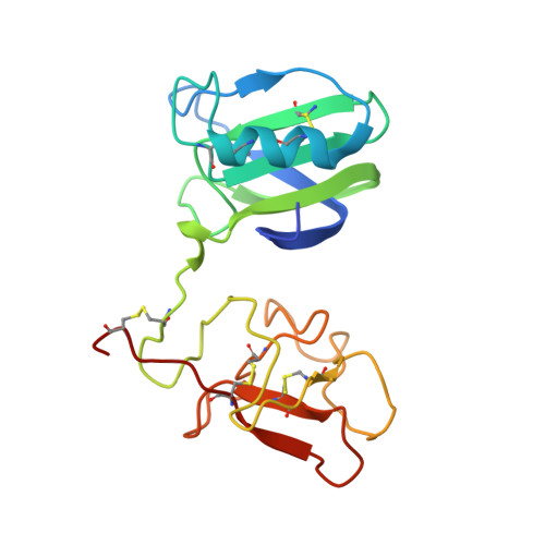
    NK1 is a splice variant of the polypeptide growth factor HGF/SF that consists of the N terminal (N) and first kringle (K) domains and retains receptor binding and signalling. While NK1 behaves as a monomer in solution, two independent crystallographic structures have previously shown an identical, tightly packed dimer. Here we report a novel orthorhombic crystal form of NK1 at 2.5 A resolution in which four NK1 protomers are packed in two distinct dimers in the asymmetric unit. Although the basic architecture of the new NK1 dimers is similar to the two described earlier, the new crystal form demonstrates extensive hinge movement between the N and K domain that leads to re-orientation of the receptor-binding sites. The hinge bending is evidence of the paucity of strong interactions between domains within the protomer, in contrast to the extensive interactions between protomers in the dimer. These observations are consistent with domain swapping in the dimer, such that the interdomain interactions of the monomer are replaced by equivalent interprotomer interactions in the dimer and offer a route for protein engineering of NK1 variants which may act as receptor antagonists.


  • Organizational Affiliation

    Department of Biochemistry, University of Cambridge, 80 Tennis Court Road, Cambridge CB2 1GA, UK.


Macromolecules
Find similar proteins by:  (by identity cutoff)  |  3D Structure
Entity ID: 1
MoleculeChains Sequence LengthOrganismDetailsImage
HEPATOCYTE GROWTH FACTOR
A, B, C, D
177Homo sapiensMutation(s): 0 
UniProt & NIH Common Fund Data Resources
Find proteins for P14210 (Homo sapiens)
Explore P14210 
Go to UniProtKB:  P14210
PHAROS:  P14210
GTEx:  ENSG00000019991 
Entity Groups  
Sequence Clusters30% Identity50% Identity70% Identity90% Identity95% Identity100% Identity
UniProt GroupP14210
Sequence Annotations
Expand
  • Reference Sequence
Experimental Data & Validation

Experimental Data

  • Method: X-RAY DIFFRACTION
  • Resolution: 2.50 Å
  • R-Value Free: 0.288 
  • R-Value Work: 0.237 
  • R-Value Observed: 0.237 
  • Space Group: C 2 2 21
Unit Cell:
Length ( Å )Angle ( ˚ )
a = 73.48α = 90
b = 78.26β = 90
c = 304.46γ = 90
Software Package:
Software NamePurpose
CNSrefinement
DENZOdata reduction
SCALEPACKdata scaling
AMoREphasing

Structure Validation

View Full Validation Report


Entry History 

Deposition Data

Revision History  (Full details and data files)

  • Version 1.0: 2001-11-19
    Type: Initial release
  • Version 1.1: 2011-05-08
    Changes: Version format compliance
  • Version 1.2: 2011-07-13
    Changes: Version format compliance
  • Version 2.0: 2017-11-08
    Changes: Atomic model, Source and taxonomy, Structure summary
  • Version 2.1: 2023-12-13
    Changes: Data collection, Database references, Other, Refinement description