1E24

LYSYL-TRNA SYNTHETASE (LYSU) HEXAGONAL FORM complexed with lysine and ATP and MN2+


Experimental Data Snapshot

  • Method: X-RAY DIFFRACTION
  • Resolution: 2.35 Å
  • R-Value Free: 0.249 
  • R-Value Work: 0.182 
  • R-Value Observed: 0.182 

wwPDB Validation   3D Report Full Report


Ligand Structure Quality Assessment 


This is version 1.1 of the entry. See complete history


Literature

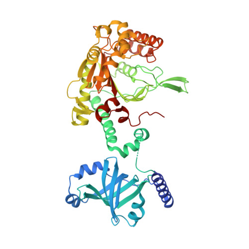
Active Site of Lysyl-tRNA Synthetase: Structural Studies of the Adenylation Reaction

Desogus, G.Todone, F.Brick, P.Onesti, S.

(2000) Biochemistry 39: 8418

  • DOI: https://doi.org/10.1021/bi0006722
  • Primary Citation of Related Structures:  
    1E1O, 1E1T, 1E22, 1E24

  • PubMed Abstract: 

    Aminoacyl-tRNA synthetases play a key role in protein biosynthesis by catalyzing the specific aminoacylation of tRNA. The energy required for the formation of the ester bond between the amino acid carboxylate group and the tRNA acceptor stem is supplied by coupling the reaction to the hydrolysis of ATP. Lysyl-tRNA synthetase from Escherichia coli belongs to the family of class II synthetases and carries out a two-step reaction, in which lysine is activated by being attached to the alpha-phosphate of AMP before being transferred to the cognate tRNA. Crystals of the thermo-inducible E. coli lysyl-tRNA synthetase LysU which diffract to 2.1 A resolution have been used to determine crystal structures of the enzyme in the presence of lysine, the lysyl-adenylate intermediate, and the nonhydrolyzable ATP analogue AMP-PCP. Additional data have been obtained from crystals soaked in a solution containing ATP and Mn(2+). The refined crystal structures give "snapshots" of the active site corresponding to key steps in the aminoacylation reaction and provide the structural framework for understanding the mechanism of lysine activation. The active site of LysU is shaped to position the substrates for the nucleophilic attack of the lysine carboxylate on the ATP alpha-phosphate. No residues are directly involved in catalysis, but a number of highly conserved amino acids and three metal ions coordinate the substrates and stabilize the pentavalent transition state. A loop close to the catalytic pocket, disordered in the lysine-bound structure, becomes ordered upon adenine binding.


  • Organizational Affiliation

    Biophysics Section, Blackett Laboratory, Imperial College of Science, Technology and Medicine, London, UK.


Macromolecules
Find similar proteins by:  (by identity cutoff)  |  3D Structure
Entity ID: 1
MoleculeChains Sequence LengthOrganismDetailsImage
LYSYL-TRNA SYNTHETASE504Escherichia coli K-12Mutation(s): 0 
Gene Names: LYSU
EC: 6.1.1.6
UniProt
Find proteins for P0A8N5 (Escherichia coli (strain K12))
Explore P0A8N5 
Go to UniProtKB:  P0A8N5
Entity Groups  
Sequence Clusters30% Identity50% Identity70% Identity90% Identity95% Identity100% Identity
UniProt GroupP0A8N5
Sequence Annotations
Expand
  • Reference Sequence
Small Molecules
Ligands 4 Unique
IDChains Name / Formula / InChI Key2D Diagram3D Interactions
ATP
Query on ATP

Download Ideal Coordinates CCD File 
C [auth A]ADENOSINE-5'-TRIPHOSPHATE
C10 H16 N5 O13 P3
ZKHQWZAMYRWXGA-KQYNXXCUSA-N
LYS
Query on LYS

Download Ideal Coordinates CCD File 
B [auth A]LYSINE
C6 H15 N2 O2
KDXKERNSBIXSRK-YFKPBYRVSA-O
GOL
Query on GOL

Download Ideal Coordinates CCD File 
G [auth A],
H [auth A]
GLYCEROL
C3 H8 O3
PEDCQBHIVMGVHV-UHFFFAOYSA-N
MN
Query on MN

Download Ideal Coordinates CCD File 
D [auth A],
E [auth A],
F [auth A]
MANGANESE (II) ION
Mn
WAEMQWOKJMHJLA-UHFFFAOYSA-N
Experimental Data & Validation

Experimental Data

  • Method: X-RAY DIFFRACTION
  • Resolution: 2.35 Å
  • R-Value Free: 0.249 
  • R-Value Work: 0.182 
  • R-Value Observed: 0.182 
  • Space Group: P 61 2 2
Unit Cell:
Length ( Å )Angle ( ˚ )
a = 143.6α = 90
b = 143.6β = 90
c = 177.23γ = 120
Software Package:
Software NamePurpose
X-PLORrefinement
MOSFLMdata reduction
CCP4data scaling
X-PLORphasing

Structure Validation

View Full Validation Report


Ligand Structure Quality Assessment 


Entry History 

Deposition Data

Revision History  (Full details and data files)

  • Version 1.0: 2000-07-28
    Type: Initial release
  • Version 1.1: 2011-08-24
    Changes: Atomic model, Derived calculations, Non-polymer description, Other, Refinement description, Version format compliance