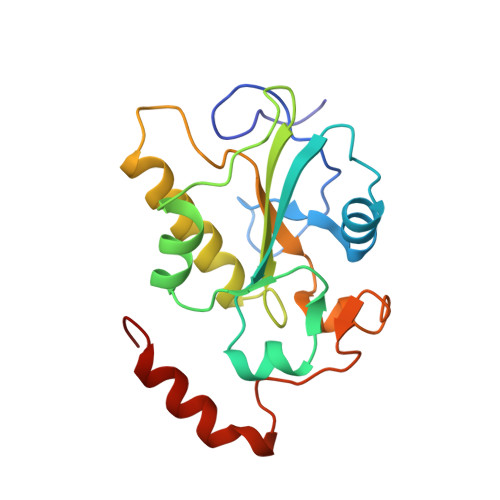
Crystal structure of the catalytic subunit of Cdc25B required for G2/M phase transition of the cell cycle.
Reynolds, R.A., Yem, A.W., Wolfe, C.L., Deibel Jr., M.R., Chidester, C.G., Watenpaugh, K.D.(1999) J Mol Biol 293: 559-568
- PubMed: 10543950
- DOI: https://doi.org/10.1006/jmbi.1999.3168
- Primary Citation of Related Structures:
1CWR, 1CWS, 1CWT, 1QB0 - PubMed Abstract:
Cdc25B is a dual specificity phosphatase involved in the control of cyclin-dependent kinases and the progression of cells through the cell cycle. A series of minimal domain Cdc25B constructs maintaining catalytic activity have been expressed. The structure of a minimum domain construct binding sulfate was determined at 1.9 A resolution and a temperature of 100 K. Other forms of the same co?nstruct were determined at lower resolution and room temperature. The overall folding and structure of the domain is similar to that found for Cdc25A. An important difference between the two is that the Cdc25B domain binds oxyanions in the catalytic site while that of Cdc25A appears unable to bind oxyanions. There are also important conformational differences in the C-terminal region. In Cdc25B, both sulfate and tungstate anions are shown to bind in the catalytic site containing the signature motif (HCxxxxxR) in a conformation similar to that of other protein tyrosine phosphatases and dual specificity phosphatases, with the exception of the Cdc25A. The Cdc25B constructs, with various truncations of the C-terminal residues, are shown to have potent catalytic activity. When cut back to the site at which the Cdc25A structure begins to deviate from the Cdc25B structure, the activity is considerably less. There is a pocket extending from the catalytic site to an anion-binding site containing a chloride about 14 A away. The catalytic cysteine residue, Cys473, can be oxidized to form a disulfide linkage to Cys426. A readily modifiable cysteine residue, Cys484, resides in another pocket that binds a sulfate but not in the signature motif conformation. This region of the structure is highly conserved between the Cdc25 molecules and could serve some unknown function.
Organizational Affiliation:
Department of Physics, Grand Valley State University, Allendale, MI, USA.