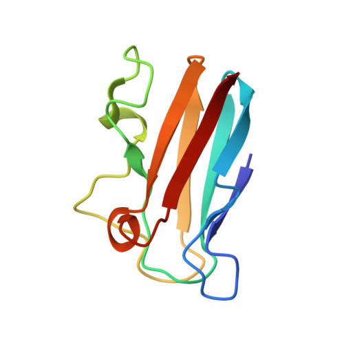
High-resolution solution structure of reduced French bean plastocyanin and comparison with the crystal structure of poplar plastocyanin.
Moore, J.M., Lepre, C.A., Gippert, G.P., Chazin, W.J., Case, D.A., Wright, P.E.(1991) J Mol Biol 221: 533-555
- PubMed: 1920431
- DOI: https://doi.org/10.1016/0022-2836(91)80071-2
- Primary Citation of Related Structures:
9PCY - PubMed Abstract:
The three-dimensional solution structure of reduced (CuI) plastocyanin from French bean leaves has been determined by distance geometry and restrained molecular dynamics methods using constraints obtained from 1H n.m.r. (nuclear magnetic resonance) spectroscopy. A total of 1244 experimental constraints were used, including 1120 distance constraints, 103 dihedral angle constraints and 21 hydrogen bond constraints. Stereospecific assignments were made for 26 methylene groups and the methyls of 11 valines. Additional constraints on copper co-ordination were included in the restrained dynamics calculations. The structures are well defined with average atomic root-mean-square deviations from the mean of 0.45 A for all backbone heavy atoms and 1.08 A for side-chain heavy atoms. French bean plastocyanin adopts a beta-sandwich structure in solution that is similar to the X-ray structure of reduced poplar plastocyanin; the average atomic root-mean-square difference between 16 n.m.r. structures and the X-ray structure is 0.76 A for all backbone heavy atoms. The conformations of the side-chains that constitute the hydrophobic core of French bean plastocyanin are very well defined. Of 47 conserved residues that populate a single chi 1 angle in solution, 43 have the same rotamer in the X-ray structure. Many surface side-chains adopt highly preferred conformations in solution, although the 3J alpha beta coupling constants often indicate some degree of conformational averaging. Some surface side-chains are disordered in both the solution and crystal structures of plastocyanin. There is a striking correlation between measures of side-chain disorder in solution and side-chain temperature factors in the X-ray structure. Side-chains that form a distinctive acidic surface region, believed to be important in binding other electron transfer proteins, appear to be disordered. Fifty backbone amide protons form hydrogen bonds to carbonyls in more than 60% of the n.m.r. structures; 45 of these amide protons exchange slowly with solvent deuterons. Ten hydrogen bonds are formed between side-chain and backbone atoms, eight of which are correlated with decreased proton exchange. Of the 60 hydrogen bonds formed in French bean plastocyanin, 56 occur in the X-ray structure of the poplar protein; two of the missing hydrogen bonds are absent as a result of mutations. It appears that molecular dynamics refinement of highly constrained n.m.r. structures allows accurate prediction of the pattern of hydrogen bonding.
Organizational Affiliation:
Department of Molecular Biology, Research Institute of Scripps Clinic, La Jolla, CA 92037.