


Crystal structure of the ternary complex of Phosphopantetheine adenylyltransferase (PPAT) from Enterobacter sp. with Coenzyme-A and Phosphonoacetic acid at 2.07 A resolution.
Ahmad, N., Sharma, P., Sharma, S., Singh, T.P.To be published.
Experimental Data Snapshot
Entity ID: 1 | |||||
|---|---|---|---|---|---|
| Molecule | Chains | Sequence Length | Organism | Details | Image |
| Phosphopantetheine adenylyltransferase | 166 | Enterobacter sp. 638 | Mutation(s): 0 Gene Names: coaD, Ent638_0105 EC: 2.7.7.3 | 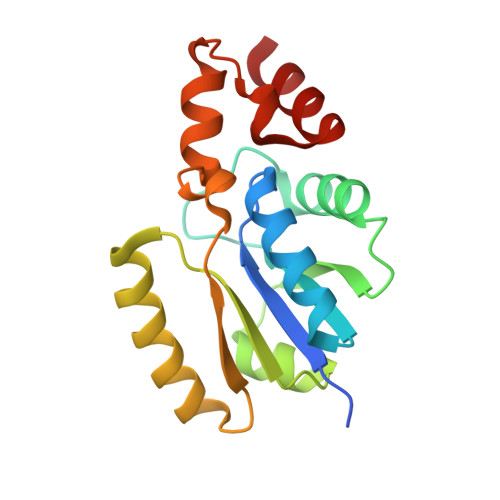 | |
UniProt | |||||
Find proteins for A4W515 (Enterobacter sp. (strain 638)) Explore A4W515 Go to UniProtKB: A4W515 | |||||
Entity Groups | |||||
| Sequence Clusters | 30% Identity50% Identity70% Identity90% Identity95% Identity100% Identity | ||||
| UniProt Group | A4W515 | ||||
Sequence AnnotationsExpand | |||||
| |||||
| Ligands 4 Unique | |||||
|---|---|---|---|---|---|
| ID | Chains | Name / Formula / InChI Key | 2D Diagram | 3D Interactions | |
| COA (Subject of Investigation/LOI) Query on COA | G [auth A], O [auth E] | COENZYME A C21 H36 N7 O16 P3 S RGJOEKWQDUBAIZ-IBOSZNHHSA-N |  | ||
| PAE (Subject of Investigation/LOI) Query on PAE | H [auth A] K [auth B] L [auth C] M [auth D] N [auth E] | PHOSPHONOACETIC ACID C2 H5 O5 P XUYJLQHKOGNDPB-UHFFFAOYSA-N |  | ||
| GOL Query on GOL | J [auth B], P [auth F] | GLYCEROL C3 H8 O3 PEDCQBHIVMGVHV-UHFFFAOYSA-N |  | ||
| EDO Query on EDO | I [auth B] | 1,2-ETHANEDIOL C2 H6 O2 LYCAIKOWRPUZTN-UHFFFAOYSA-N |  | ||
| Length ( Å ) | Angle ( ˚ ) |
|---|---|
| a = 137.966 | α = 90 |
| b = 78.659 | β = 93.121 |
| c = 107.027 | γ = 90 |
| Software Name | Purpose |
|---|---|
| REFMAC | refinement |
| MxCuBE | data collection |
| XDS | data reduction |
| Aimless | data scaling |
| MOLREP | phasing |
| Coot | model building |
| Funding Organization | Location | Grant Number |
|---|---|---|
| Indian Council of Medical Research | India | I-1251 |
RCSB PDB (citation) is hosted by
RCSB PDB is a member of the

