8HVP

STRUCTURE AT 2.5-ANGSTROMS RESOLUTION OF CHEMICALLY SYNTHESIZED HUMAN IMMUNODEFICIENCY VIRUS TYPE 1 PROTEASE COMPLEXED WITH A HYDROXYETHYLENE*-BASED INHIBITOR


Experimental Data Snapshot

  • Method: X-RAY DIFFRACTION
  • Resolution: 2.50 Å
  • R-Value Observed: 0.138 

wwPDB Validation   3D Report Full Report


This is version 2.0 of the entry. See complete history


Literature

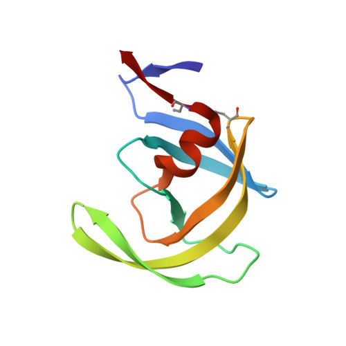
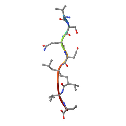
Structure at 2.5-A resolution of chemically synthesized human immunodeficiency virus type 1 protease complexed with a hydroxyethylene-based inhibitor.

Jaskolski, M.Tomasselli, A.G.Sawyer, T.K.Staples, D.G.Heinrikson, R.L.Schneider, J.Kent, S.B.Wlodawer, A.

(1991) Biochemistry 30: 1600-1609

  • DOI: https://doi.org/10.1021/bi00220a023
  • Primary Citation of Related Structures:  
    8HVP

  • PubMed Abstract: 

    The crystal structure of a complex between chemically synthesized human immunodeficiency virus type 1 (HIV-1) protease and an octapeptide inhibitor has been refined to an R factor of 0.138 at 2.5-A resolution. The substrate-based inhibitor, H-Val-Ser-Gln-Asn-Leu psi [CH(OH)CH2]Val-Ile-Val-OH (U-85548e) contains a hydroxyethylene isostere replacement at the scissile bond that is believed to mimic the tetrahedral transition state of the proteolytic reaction. This potent inhibitor has Ki less than 1 nM and was developed as an active-site titrant of the HIV-1 protease. The inhibitor binds in an extended conformation and is involved in beta-sheet interactions with the active-site floor and flaps of the enzyme, which form the substrate/inhibitor cavity. The inhibitor diastereomer has the S configuration at the chiral carbon atom of the hydroxyethylene insert, and the hydroxyl group is within H-bonding distance of the two active-site carboxyl groups in the enzyme dimer. The two subunits of the enzyme are related by a pseudodyad, which superposes them at a 178 degrees rotation. The main difference between the subunits is in the beta turns of the flaps, which have different conformations in the two monomers. The inhibitor has a clear preferred orientation in the active site and the alternative conformation, if any, is a minor one (occupancy of less than 30%). A new model of the enzymatic mechanism is proposed in which the proteolytic reaction is viewed as a one-step process during which the nucleophile (water molecule) and electrophile (an acidic proton) attack the scissile bond in a concerted manner.


  • Organizational Affiliation

    Macromolecular Structure Laboratory, National Cancer Institute-Frederick Cancer Research and Development Center, Maryland 21702-1201.


Macromolecules
Find similar proteins by:  (by identity cutoff)  |  3D Structure
Entity ID: 1
MoleculeChains Sequence LengthOrganismDetailsImage
HIV-1 PROTEASE
A, B
99Human immunodeficiency virus 1Mutation(s): 0 
UniProt
Find proteins for P03369 (Human immunodeficiency virus type 1 group M subtype B (isolate ARV2/SF2))
Explore P03369 
Go to UniProtKB:  P03369
Entity Groups  
Sequence Clusters30% Identity50% Identity70% Identity90% Identity95% Identity100% Identity
UniProt GroupP03369
Sequence Annotations
Expand
  • Reference Sequence

Find similar proteins by:  Sequence   |   3D Structure  

Entity ID: 2
MoleculeChains Sequence LengthOrganismDetailsImage
INHIBITOR VAL-SER-GLN-ASN-LEU-PSI(CH(OH)-CH2)-VAL-ILE-VAL (U-85548E)C [auth I]7N/AMutation(s): 0 
Sequence Annotations
Expand
  • Reference Sequence
Small Molecules
Modified Residues  1 Unique
IDChains TypeFormula2D DiagramParent
ABA
Query on ABA
A, B
L-PEPTIDE LINKINGC4 H9 N O2ALA
Biologically Interesting Molecules (External Reference) 1 Unique
Experimental Data & Validation

Experimental Data

  • Method: X-RAY DIFFRACTION
  • Resolution: 2.50 Å
  • R-Value Observed: 0.138 
  • Space Group: P 21 21 21
Unit Cell:
Length ( Å )Angle ( ˚ )
a = 51.65α = 90
b = 58.76β = 90
c = 61.62γ = 90
Software Package:
Software NamePurpose
PROFFTrefinement

Structure Validation

View Full Validation Report


Entry History 

Deposition Data

Revision History  (Full details and data files)

  • Version 1.0: 1993-10-31
    Type: Initial release
  • Version 1.1: 2008-03-03
    Changes: Version format compliance
  • Version 1.2: 2011-07-13
    Changes: Atomic model, Database references, Derived calculations, Non-polymer description, Structure summary, Version format compliance
  • Version 1.3: 2012-12-12
    Changes: Other
  • Version 1.4: 2017-11-29
    Changes: Derived calculations, Other
  • Version 2.0: 2023-11-15
    Changes: Atomic model, Data collection, Database references, Derived calculations