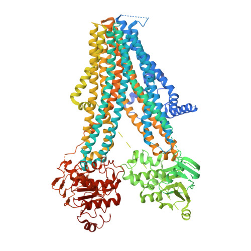
7WIT

Structure of SUR1 in complex with mitiglinide


Experimental Data Snapshot

  • Method: ELECTRON MICROSCOPY
  • Resolution: 3.21 Å
  • Aggregation State: PARTICLE 
  • Reconstruction Method: SINGLE PARTICLE 

wwPDB Validation   3D Report Full Report


This is version 1.1 of the entry. See complete history


Literature

Structural Insights Into the High Selectivity of the Anti-Diabetic Drug Mitiglinide

Wang, M.M.Wu, J.X.Chen, L.

(2022) Front Pharmacol 13: 929684-929684

  • DOI: https://doi.org/10.3389/fphar.2022.929684
  • Primary Citation of Related Structures:  
    7WIT

  • PubMed Abstract: 

    Mitiglinide is a highly selective fast-acting anti-diabetic drug that induces insulin secretion by inhibiting pancreatic K ATP channels. However, how mitiglinide binds K ATP channels remains unknown. Here, we show the cryo-EM structure of the SUR1 subunit complexed with mitiglinide. The structure reveals that mitiglinide binds inside the common insulin secretagogue-binding site of SUR1, which is surrounded by TM7, TM8, TM16, and TM17. Mitiglinide locks SUR1 in the NBD-separated inward-facing conformation. The detailed structural analysis of the mitiglinide-binding site uncovers the molecular basis of its high selectivity.


  • Organizational Affiliation

    State Key Laboratory of Membrane Biology, Beijing Key Laboratory of Cardiometabolic Molecular Medicine, College of Future Technology, Institute of Molecular Medicine, Peking University, Beijing, China.


Macromolecules
Find similar proteins by:  (by identity cutoff)  |  3D Structure
Entity ID: 1
MoleculeChains Sequence LengthOrganismDetailsImage
ATP-sensitive inward rectifier potassium channel 11,ATP-binding cassette sub-family C member 8 isoform X11,434Bos taurusMesocricetus auratus
This entity is chimeric
Mutation(s): 0 
Gene Names: KCNJ11Abcc8
Membrane Entity: Yes 
UniProt
Find proteins for A2VDS4 (Bos taurus)
Explore A2VDS4 
Go to UniProtKB:  A2VDS4
Find proteins for A0A1U7R319 (Mesocricetus auratus)
Explore A0A1U7R319 
Go to UniProtKB:  A0A1U7R319
Entity Groups  
Sequence Clusters30% Identity50% Identity70% Identity90% Identity95% Identity100% Identity
UniProt GroupsA0A1U7R319A2VDS4
Sequence Annotations
Expand
  • Reference Sequence
Small Molecules
Ligands 2 Unique
IDChains Name / Formula / InChI Key2D Diagram3D Interactions
ATP
Query on ATP

Download Ideal Coordinates CCD File 
B [auth A]ADENOSINE-5'-TRIPHOSPHATE
C10 H16 N5 O13 P3
ZKHQWZAMYRWXGA-KQYNXXCUSA-N
9I0 (Subject of Investigation/LOI)
Query on 9I0

Download Ideal Coordinates CCD File 
C [auth A](2S)-4-[(3aR,7aS)-1,3,3a,4,5,6,7,7a-octahydroisoindol-2-yl]-4-oxidanylidene-2-(phenylmethyl)butanoic acid
C19 H25 N O3
WPGGHFDDFPHPOB-BBWFWOEESA-N
Experimental Data & Validation

Experimental Data

  • Method: ELECTRON MICROSCOPY
  • Resolution: 3.21 Å
  • Aggregation State: PARTICLE 
  • Reconstruction Method: SINGLE PARTICLE 

Structure Validation

View Full Validation Report


Entry History & Funding Information

Deposition Data


Funding OrganizationLocationGrant Number
National Natural Science Foundation of China (NSFC)China91957201 and 31870833

Revision History  (Full details and data files)

  • Version 1.0: 2022-11-16
    Type: Initial release
  • Version 1.1: 2022-12-21
    Changes: Database references