Evidence for the Chemical Mechanism of RibB (3,4-Dihydroxy-2-butanone 4-phosphate Synthase) of Riboflavin Biosynthesis.
Kenjic, N., Meneely, K.M., Wherritt, D.J., Denler, M.C., Jackson, T.A., Moran, G.R., Lamb, A.L.(2022) J Am Chem Soc 144: 12769-12780
- PubMed: 35802469
- DOI: https://doi.org/10.1021/jacs.2c03376
- Primary Citation of Related Structures:
7UEZ, 7UF0, 7UF1, 7UF2, 7UF3, 7UF4, 7UF5 - PubMed Abstract:
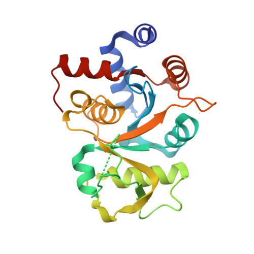
RibB (3,4-dihydroxy-2-butanone 4-phosphate synthase) is a magnesium-dependent enzyme that excises the C4 of d-ribulose-5-phosphate (d-Ru5P) as formate. RibB generates the four-carbon substrate for lumazine synthase that is incorporated into the xylene moiety of lumazine and ultimately the riboflavin isoalloxazine. The reaction was first identified by Bacher and co-workers in the 1990s, and their chemical mechanism hypothesis became canonical despite minimal direct evidence. X-ray crystal structures of RibB typically show two metal ions when solved in the presence of non-native metals and/or liganding non-substrate analogues, and the consensus hypothetical mechanism has incorporated this cofactor set. We have used a variety of biochemical approaches to further characterize the chemistry catalyzed by RibB from Vibrio cholera (VcRibB). We show that full activity is achieved at metal ion concentrations equal to the enzyme concentration. This was confirmed by electron paramagnetic resonance of the enzyme reconstituted with manganese and crystal structures liganded with Mn 2+ and a variety of sugar phosphates. Two transient species prior to the formation of products were identified using acid quench of single turnover reactions in combination with NMR for singly and fully 13 C-labeled d-Ru5P. These data indicate that dehydration of C1 forms the first transient species, which undergoes rearrangement by a 1,2 migration, fusing C5 to C3 and generating a hydrated C4 that is poised for elimination as formate. Structures determined from time-dependent Mn 2+ soaks of VcRibB-d-Ru5P crystals show accumulation in crystallo of the same intermediates. Collectively, these data reveal for the first time crucial transient chemical states in the mechanism of RibB.
Organizational Affiliation:
Department of Molecular Biosciences, University of Kansas, Lawrence, Kansas 66045, United States.