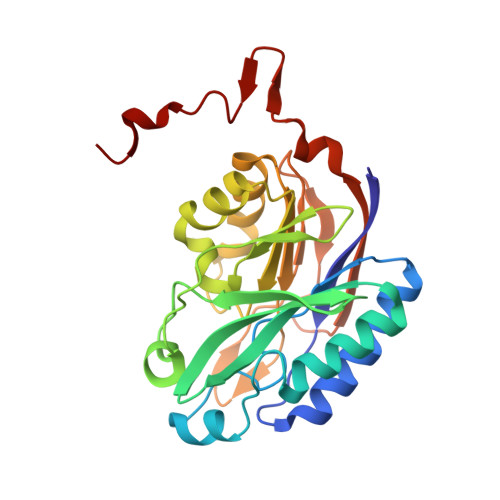
The structures of the C146A variant of the amidase from Pyrococcus horikoshii bound to glutaramide and acetamide suggest the basis of amide recognition.
Makumire, S., Su, S., Weber, B.W., Woodward, J.D., Wangari Kimani, S., Hunter, R., Sewell, B.T.(2022) J Struct Biol 214: 107859-107859
- PubMed: 35439644
- DOI: https://doi.org/10.1016/j.jsb.2022.107859
- Primary Citation of Related Structures:
7OVG - PubMed Abstract:
The nitrilase superfamily enzymes from Pyrococcus abyssi and Pyrococcus horikoshii hydrolyze several different amides. No nitriles that we tested were hydrolyzed by either enzyme. Propionamide and acetamide were the most rapidly hydrolyzed of all the substrates tested. Amide substrate docking studies on the wild-type and C146A variant P. horikoshii enzymes suggest a sequence in which the incoming amide substrate initially hydrogen bonds to the amino group of Lys-113 and the backbone carbonyl of Asn-171. When steric hindrance is relieved by replacing the cysteine with alanine, the amide then docks such that the amino group of Lys-113 and the backbone amide of Phe-147 are hydrogen-bonded to the substrate carbonyl oxygen, while the backbone carbonyl oxygen of Asn-171 and the carboxyl oxygen of Glu-42 are hydrogen-bonded to the amino group of the substrate. Here, we confirm the location of the acetamide and glutaramide ligands experimentally in well-resolved crystal structures of the C146A mutant of the enzyme from P. horikoshii. This ligand location suggests that there is no direct interaction between the substrate amide and the other active site glutamate, Glu-120, and supports an active-site geometry leading to the formation of the thioester intermediate via an attack on the si-face of the amide by the sulfhydryl of the active site cysteine.
Organizational Affiliation:
Structural Biology Research Unit, Department of Integrative Biomedical Sciences, University of Cape Town, Observatory 7925, South Africa. Electronic address: stanmakster@gmail.com.