7L7X

X-ray structure of the Pcryo_0638 aminotransferase from Psychrobacter cryohalolentis


Experimental Data Snapshot

  • Method: X-RAY DIFFRACTION
  • Resolution: 1.30 Å
  • R-Value Free: 0.169 
  • R-Value Work: 0.150 

wwPDB Validation   3D Report Full Report


Ligand Structure Quality Assessment 


This is version 1.2 of the entry. See complete history


Literature

Characterization of two enzymes from Psychrobacter cryohalolentis that are required for the biosynthesis of an unusual diacetamido-d-sugar.

Linehan, M.P.Thoden, J.B.Holden, H.M.

(2021) J Biol Chem 296: 100463-100463

  • DOI: https://doi.org/10.1016/j.jbc.2021.100463
  • Primary Citation of Related Structures:  
    7L7X, 7L7Y, 7L7Z, 7L81, 7L82

  • PubMed Abstract: 

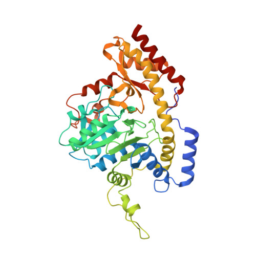
    Psychrobacter cryohalolentis strain K5 T is a Gram-negative organism first isolated in 2006. It has a complex O-antigen that contains, in addition to l-rhamnose and d-galactose, two diacetamido- and a triacetamido-sugar. The biochemical pathways for the production of these unusual sugars are presently unknown. Utilizing the published genome sequence of the organism, we hypothesized that the genes 0612, 0638, and 0637 encode for a 4,6-dehydratase, an aminotransferase, and an N-acetyltransferase, respectively, which would be required for the biosynthesis of one of the diacetamido-sugars, 2,4-diacetamido-2,4,6-trideoxy-d-glucose, starting from UDP-N-acetylglucosamine. Here we present functional and structural data on the proteins encoded by the 0638 and 0637 genes. The kinetic properties of these enzymes were investigated by a discontinuous HPLC assay. An X-ray crystallographic structure of 0638, determined in its external aldimine form to 1.3 Å resolution, demonstrated the manner in which the UDP ligand is positioned into the active site. It is strikingly different from that previously observed for PglE from Campylobacter jejuni, which functions on the same substrate. Four X-ray crystallographic structures were also determined for 0637 in various complexed states at resolutions between 1.3 and 1.55 Å. Remarkably, a tetrahedral intermediate mimicking the presumed transition state was trapped in one of the complexes. The data presented herein confirm the hypothesized functions of these enzymes and provide new insight into an unusual sugar biosynthetic pathway in Gram-negative bacteria. We also describe an efficient method for acetyl-CoA synthesis that allowed us to overcome its prohibitive cost for this analysis.


  • Organizational Affiliation

    Department of Biochemistry, University of Wisconsin, Madison, Wisconsin, USA.


Macromolecules
Find similar proteins by:  (by identity cutoff)  |  3D Structure
Entity ID: 1
MoleculeChains Sequence LengthOrganismDetailsImage
DegT/DnrJ/EryC1/StrS aminotransferaseA [auth AAA],
B [auth BBB]
404Psychrobacter cryohalolentis K5Mutation(s): 0 
Gene Names: Pcryo_0638
UniProt
Find proteins for Q1QD32 (Psychrobacter cryohalolentis (strain ATCC BAA-1226 / DSM 17306 / VKM B-2378 / K5))
Explore Q1QD32 
Go to UniProtKB:  Q1QD32
Entity Groups  
Sequence Clusters30% Identity50% Identity70% Identity90% Identity95% Identity100% Identity
UniProt GroupQ1QD32
Sequence Annotations
Expand
  • Reference Sequence
Small Molecules
Ligands 5 Unique
IDChains Name / Formula / InChI Key2D Diagram3D Interactions
4RA (Subject of Investigation/LOI)
Query on 4RA

Download Ideal Coordinates CCD File 
D [auth AAA],
N [auth BBB]
[(2R,3R,4R,5S,6R)-3-acetamido-6-methyl-5-[(E)-[2-methyl-3-oxidanyl-5-(phosphonooxymethyl)pyridin-4-yl]methylideneamino]-4-oxidanyl-oxan-2-yl] [[(2R,3S,4R,5R)-5-[2,4-bis(oxidanylidene)pyrimidin-1-yl]-3,4-bis(oxidanyl)oxolan-2-yl]methoxy-oxidanyl-phosphoryl] hydrogen phosphate
C25 H36 N5 O20 P3
BKHPSAUTHKIVAK-OTBUILFPSA-N
PMP
Query on PMP

Download Ideal Coordinates CCD File 
C [auth AAA],
M [auth BBB]
4'-DEOXY-4'-AMINOPYRIDOXAL-5'-PHOSPHATE
C8 H13 N2 O5 P
ZMJGSOSNSPKHNH-UHFFFAOYSA-N
TMA
Query on TMA

Download Ideal Coordinates CCD File 
J [auth AAA],
T [auth BBB]
TETRAMETHYLAMMONIUM ION
C4 H12 N
QEMXHQIAXOOASZ-UHFFFAOYSA-N
EDO
Query on EDO

Download Ideal Coordinates CCD File 
E [auth AAA]
F [auth AAA]
G [auth AAA]
H [auth AAA]
I [auth AAA]
E [auth AAA],
F [auth AAA],
G [auth AAA],
H [auth AAA],
I [auth AAA],
O [auth BBB],
P [auth BBB],
Q [auth BBB],
R [auth BBB],
S [auth BBB]
1,2-ETHANEDIOL
C2 H6 O2
LYCAIKOWRPUZTN-UHFFFAOYSA-N
NA
Query on NA

Download Ideal Coordinates CCD File 
K [auth AAA],
L [auth AAA]
SODIUM ION
Na
FKNQFGJONOIPTF-UHFFFAOYSA-N
Experimental Data & Validation

Experimental Data

  • Method: X-RAY DIFFRACTION
  • Resolution: 1.30 Å
  • R-Value Free: 0.169 
  • R-Value Work: 0.150 
  • Space Group: P 1
Unit Cell:
Length ( Å )Angle ( ˚ )
a = 57.529α = 81.747
b = 63.141β = 78.593
c = 69.853γ = 66.818
Software Package:
Software NamePurpose
REFMACrefinement
SAINTdata reduction
SADABSdata scaling
PHASERphasing

Structure Validation

View Full Validation Report


Ligand Structure Quality Assessment 


Entry History & Funding Information

Deposition Data


Funding OrganizationLocationGrant Number
National Institutes of Health/National Institute of General Medical Sciences (NIH/NIGMS)United StatesGM134643

Revision History  (Full details and data files)

  • Version 1.0: 2021-03-03
    Type: Initial release
  • Version 1.1: 2021-09-15
    Changes: Database references
  • Version 1.2: 2023-10-18
    Changes: Data collection, Refinement description